Copyright
©The Author(s) 2020.
World J Hepatol. Nov 27, 2020; 12(11): 1128-1135
Published online Nov 27, 2020. doi: 10.4254/wjh.v12.i11.1128
Published online Nov 27, 2020. doi: 10.4254/wjh.v12.i11.1128
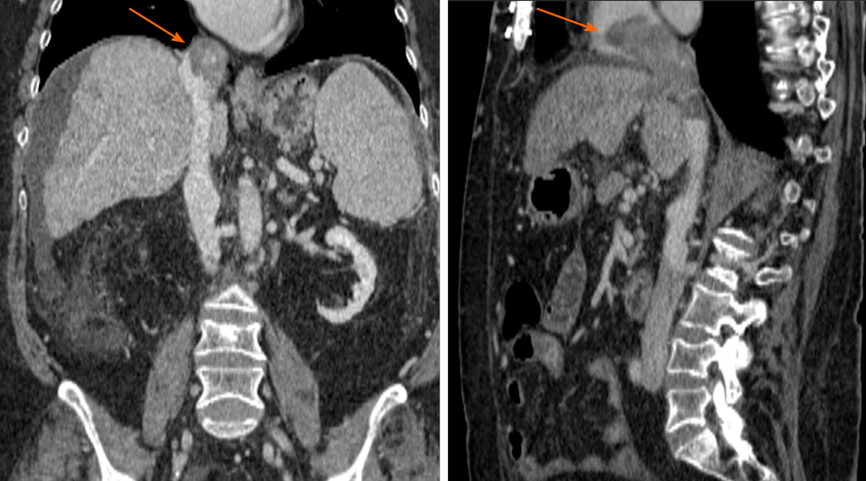
Figure 3 Computed tomography scan coronal and sagittal views show a 55 mm × 90 mm mass in segment VIII with a tumor thrombus extending to the inferior vena cava (left arrow) and reaching up to right atrium (right arrow).
Left intrahepatic portal thrombosis. Portal vein thrombosis with hypercaptation of the thrombus, suggestive of infiltrative tumor.
- Citation: Gomez-Puerto D, Mirallas O, Vidal-González J, Vargas V. Hepatocellular carcinoma with tumor thrombus extends to the right atrium and portal vein: A case report. World J Hepatol 2020; 12(11): 1128-1135
- URL: https://www.wjgnet.com/1948-5182/full/v12/i11/1128.htm
- DOI: https://dx.doi.org/10.4254/wjh.v12.i11.1128


