Copyright
©The Author(s) 2020.
World J Hepatol. Nov 27, 2020; 12(11): 1128-1135
Published online Nov 27, 2020. doi: 10.4254/wjh.v12.i11.1128
Published online Nov 27, 2020. doi: 10.4254/wjh.v12.i11.1128
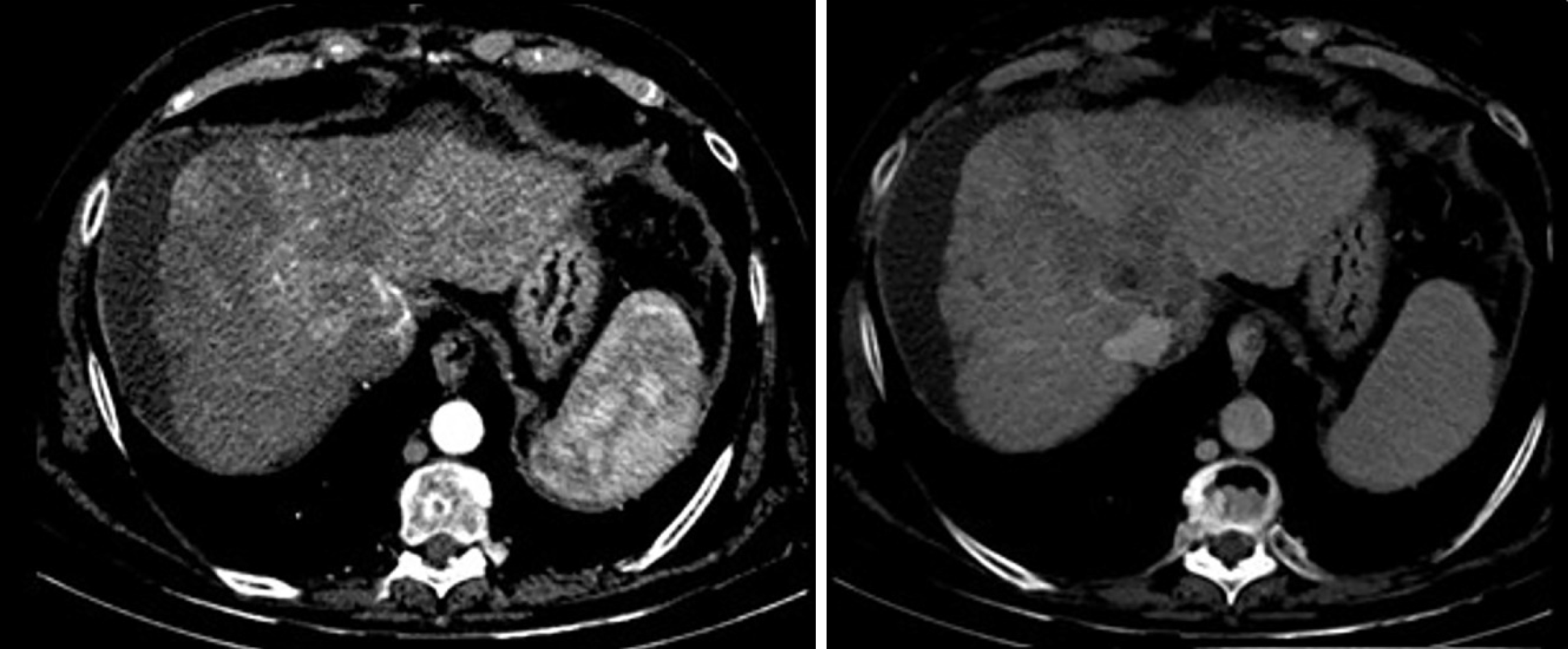
Figure 2 Computed tomography scan axial view shows a poorly defined liver injury with margins of 55 mm × 90 mm located in segment VIII and with extension to segment V and segment I, enhanced in arterial phase with fast venous phase washing.
- Citation: Gomez-Puerto D, Mirallas O, Vidal-González J, Vargas V. Hepatocellular carcinoma with tumor thrombus extends to the right atrium and portal vein: A case report. World J Hepatol 2020; 12(11): 1128-1135
- URL: https://www.wjgnet.com/1948-5182/full/v12/i11/1128.htm
- DOI: https://dx.doi.org/10.4254/wjh.v12.i11.1128


