Copyright
©The Author(s) 2020.
World J Hepatol. Nov 27, 2020; 12(11): 1128-1135
Published online Nov 27, 2020. doi: 10.4254/wjh.v12.i11.1128
Published online Nov 27, 2020. doi: 10.4254/wjh.v12.i11.1128
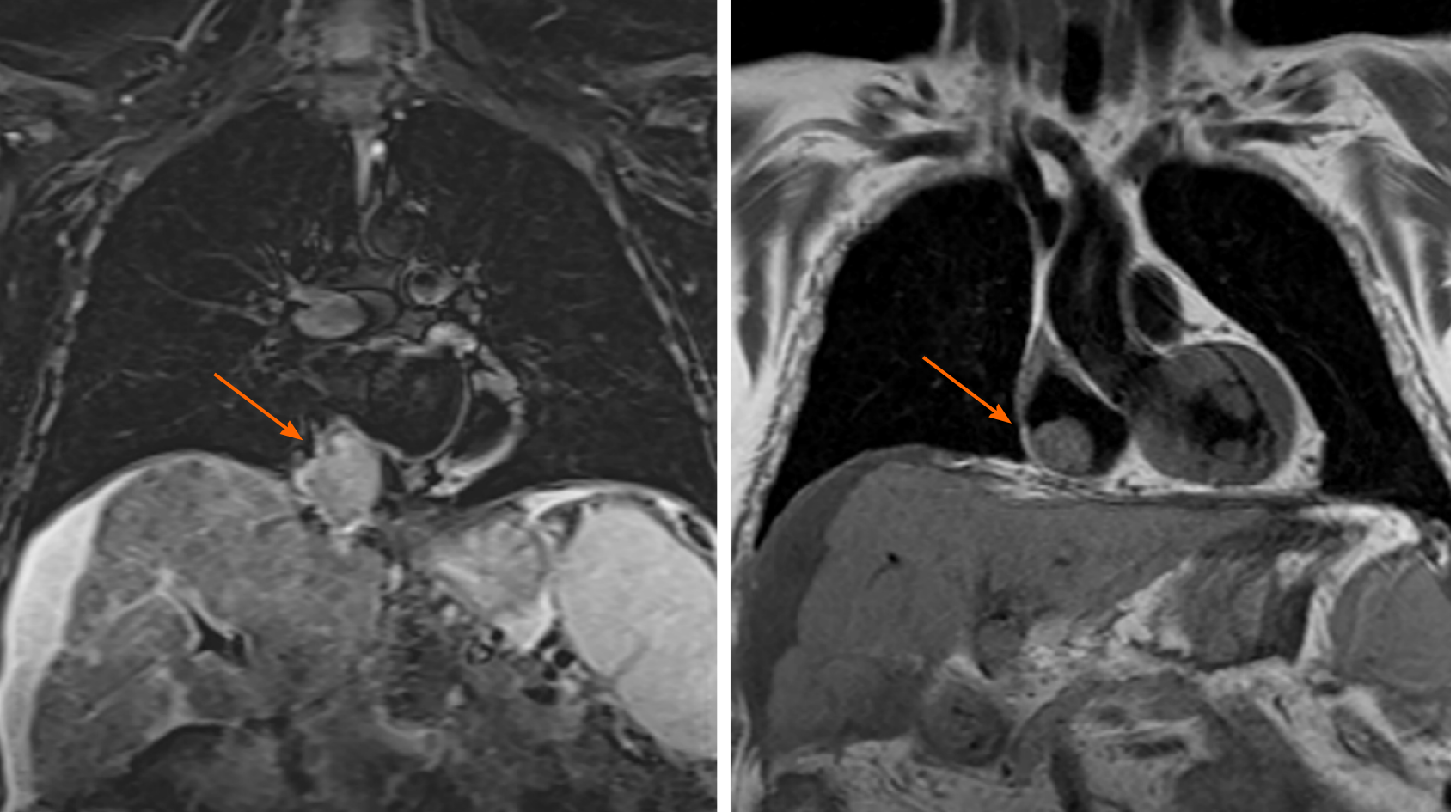
Figure 1 Magnetic resonance imaging coronal view of a polylobulated mass of 59 mm × 51 mm × 52 mm inside the right atrium (right arrow), which extends from the liver through the inferior vena cava (left arrow) to the inside of the right atrium occupying its lower-posterior wall.
These findings suggest as the first diagnostic possibility: A tumor thrombus of an advanced infiltrating hepatocellular carcinoma. Volumes and biventricular function within physiological parameters.
- Citation: Gomez-Puerto D, Mirallas O, Vidal-González J, Vargas V. Hepatocellular carcinoma with tumor thrombus extends to the right atrium and portal vein: A case report. World J Hepatol 2020; 12(11): 1128-1135
- URL: https://www.wjgnet.com/1948-5182/full/v12/i11/1128.htm
- DOI: https://dx.doi.org/10.4254/wjh.v12.i11.1128


