Copyright
©The Author(s) 2020.
World J Hepatol. Oct 27, 2020; 12(10): 775-791
Published online Oct 27, 2020. doi: 10.4254/wjh.v12.i10.775
Published online Oct 27, 2020. doi: 10.4254/wjh.v12.i10.775
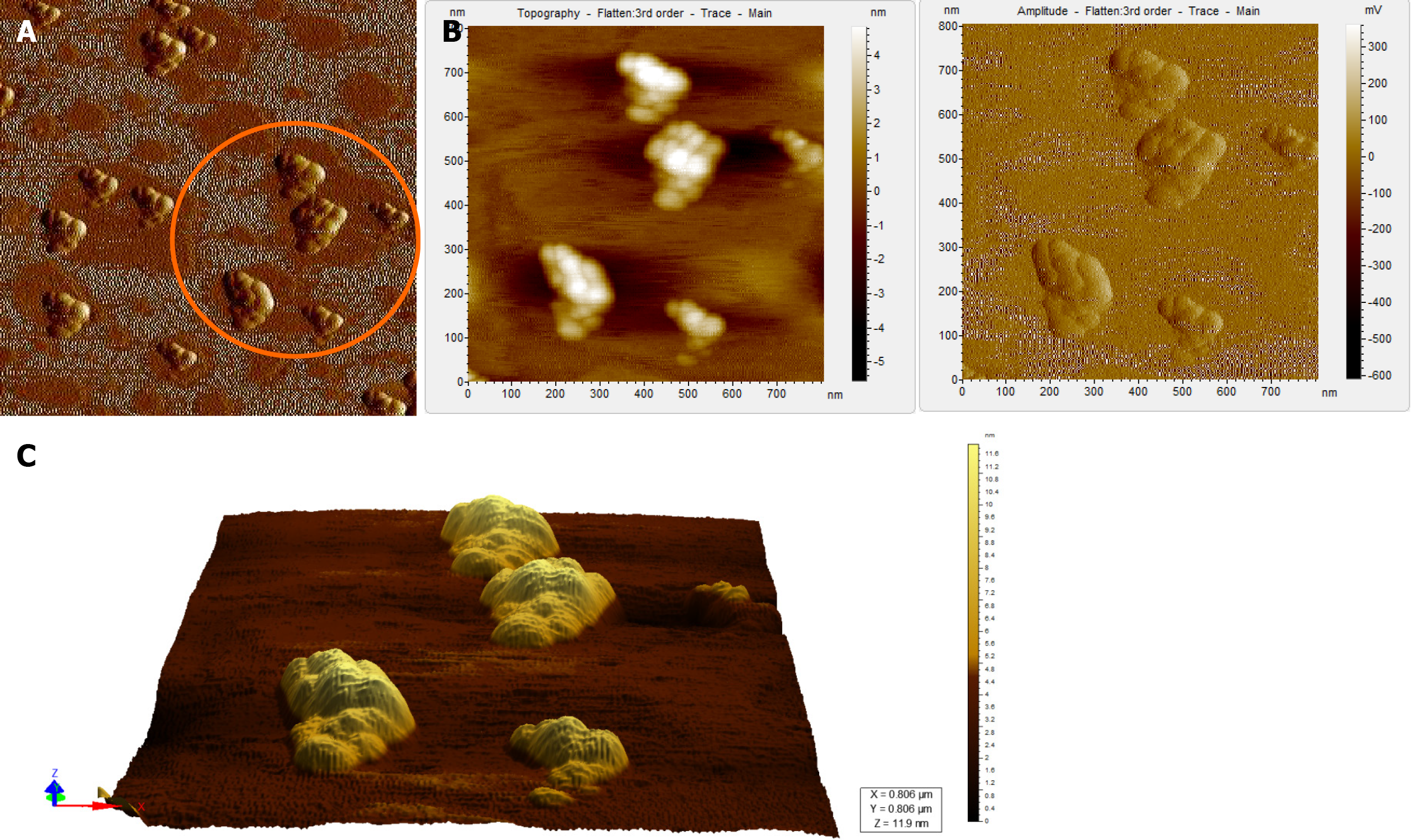
Figure 5 Atomic force microscopy images of a hepatitis B virus-negative sample.
A: Wide field of view of clumps of globular structures that look different from that seen in Figure 4A. These clumps share no resemblance with the structure of hepatitis B virus particles; B: Magnified view of the encircled region in Figure 5A in two different contrasts; C: 3D view of the clumps.
- Citation: Das P, Supekar R, Chatterjee R, Roy S, Ghosh A, Biswas S. Hepatitis B virus detected in paper currencies in a densely populated city of India: A plausible source of horizontal transmission? World J Hepatol 2020; 12(10): 775-791
- URL: https://www.wjgnet.com/1948-5182/full/v12/i10/775.htm
- DOI: https://dx.doi.org/10.4254/wjh.v12.i10.775


