Copyright
©The Author(s) 2020.
World J Hepatol. Oct 27, 2020; 12(10): 775-791
Published online Oct 27, 2020. doi: 10.4254/wjh.v12.i10.775
Published online Oct 27, 2020. doi: 10.4254/wjh.v12.i10.775
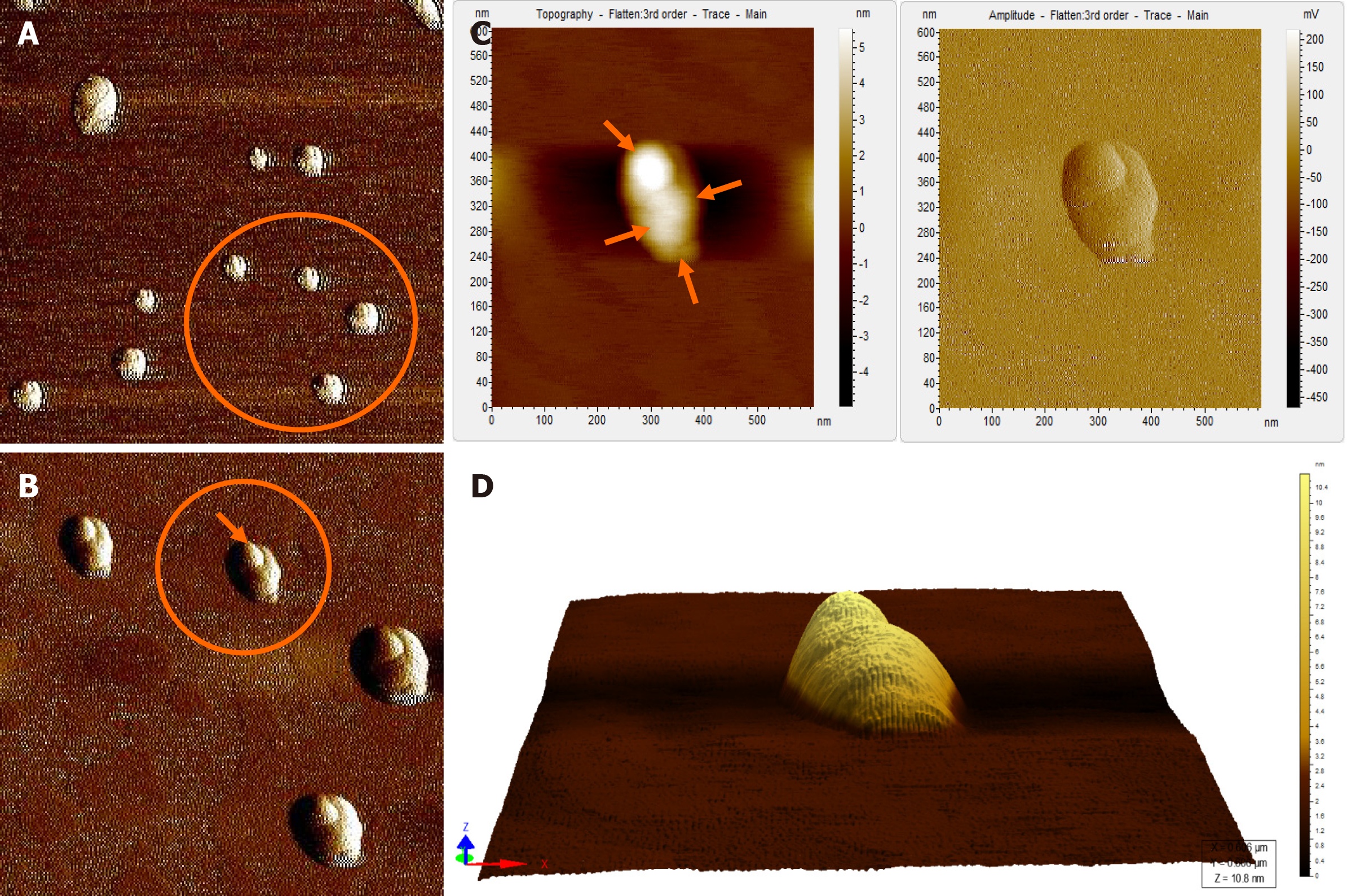
Figure 4 Atomic force microscopy images of hepatitis B virus-positive sample S8.
A: Wider field of view for the sample; B: Magnified view of the encircled area marked in Figure 4A; The encircled area illustrates representative cluster of virion particles. The arrow points to a single particle in the cluster; C: Magnified view of encircled area shown in Figure 4B in two different contrasts. The arrows show the individual intact virion particle of approximately 42 nm diameter; D: Three-dimensional (3D) view of the individual clusters seen in Figure 4C.
- Citation: Das P, Supekar R, Chatterjee R, Roy S, Ghosh A, Biswas S. Hepatitis B virus detected in paper currencies in a densely populated city of India: A plausible source of horizontal transmission? World J Hepatol 2020; 12(10): 775-791
- URL: https://www.wjgnet.com/1948-5182/full/v12/i10/775.htm
- DOI: https://dx.doi.org/10.4254/wjh.v12.i10.775


