Copyright
©The Author(s) 2020.
World J Hepatol. Jan 27, 2020; 12(1): 21-33
Published online Jan 27, 2020. doi: 10.4254/wjh.v12.i1.21
Published online Jan 27, 2020. doi: 10.4254/wjh.v12.i1.21
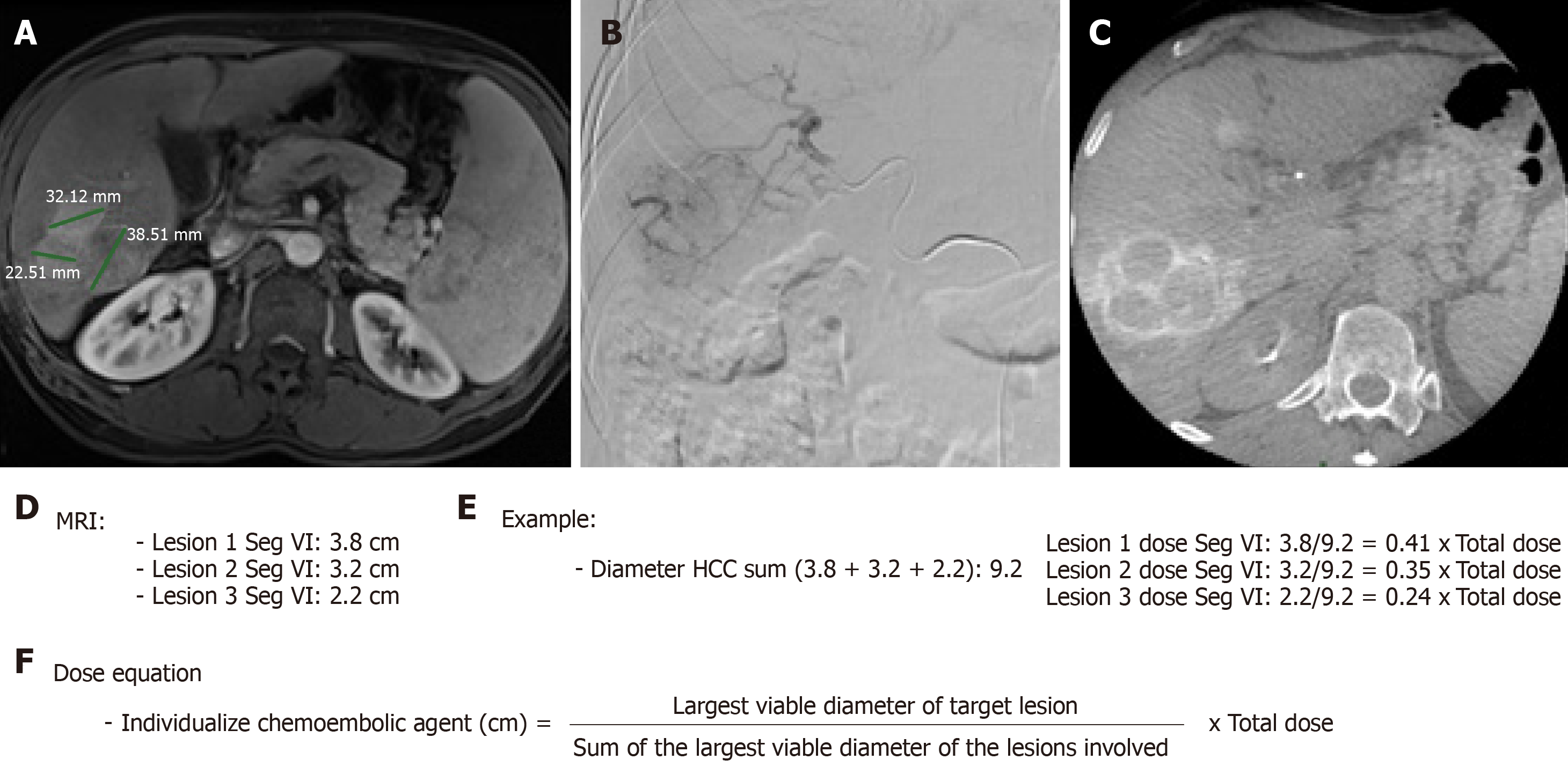
Figure 3 Calculation method for individualization of the dose of the chemoembolic agent received by treated hepatocellular carcinoma in situations of impossibility of the superselective catheterism.
A: Magnetic resonance imaging pre-chemoembolization abdomen - post-contrast T1 weighted phase - showing three confluent hypervascular lesions; B: Selective hepatic arteriography in segment VI of the right hepatic artery showing hypervascular lesions characteristic of hepatocellular carcinoma; C: Intraoperative cone beam tomography with selective arterial contrast in segment VI - venous phase - showing three confluent lesions with contrast medium lavage; D: Diameter of hepatocellular carcinomas located in segment VI to be treated; E: Exemplification of the calculation of dose individualization of the chemoembolic agent administered; F: Equation of individualized chemoembolic dose. MRI: Magnetic resonance imaging.
- Citation: Galastri FL, Nasser F, Affonso BB, Valle LGM, Odísio BC, Motta-Leal Filho JM, Salvalaggio PR, Garcia RG, de Almeida MD, Baroni RH, Wolosker N. Imaging response predictors following drug eluting beads chemoembolization in the neoadjuvant liver transplant treatment of hepatocellular carcinoma. World J Hepatol 2020; 12(1): 21-33
- URL: https://www.wjgnet.com/1948-5182/full/v12/i1/21.htm
- DOI: https://dx.doi.org/10.4254/wjh.v12.i1.21


