Copyright
©The Author(s) 2019.
World J Hepatol. May 27, 2019; 11(5): 483-488
Published online May 27, 2019. doi: 10.4254/wjh.v11.i5.483
Published online May 27, 2019. doi: 10.4254/wjh.v11.i5.483
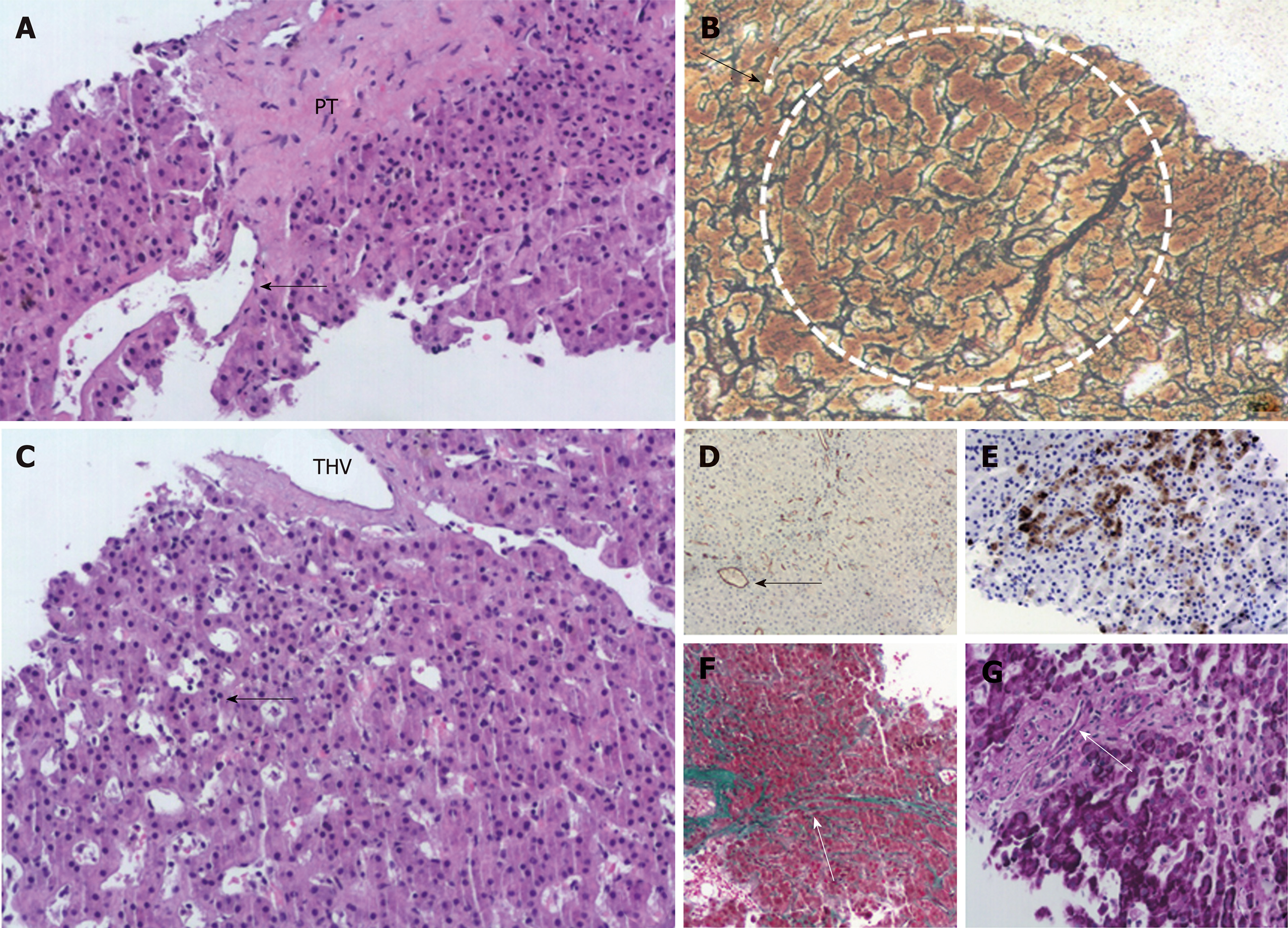
Figure 1 Selected images from the liver biopsy showing changes of portal venopathy and features of nodular regenerative hyperplasia.
A: A sclerosed PT without a visible portal vein. Peripherally, a dilated portal vein radicle herniates into the adjacent hepatic parenchyma (HE, x 10); B: Nodular regenerative hyperplasia. White dotted line circumscribes a nodule of regenerating hepatocytes surrounded by atrophic hepatocyte trabeculae (arrow), reticulin stain, x 10; C: THV with perivenular sclerosis. Sinusoidal dilatation in zone 3 (arrow), (HE, x 10); D: Sinusoidal capillarization in ischemic areas highlighted by CD34 immunostain. Arrow points to a THV (brown CD 34-positive sinusoidal staining), x 10; E: Keratin 7-positive atrophic hepatocytes indicative of ischemia in zone 3 (brown staining), x 10; F: Sinusoidal zone 3 fibrosis in zone 3 (arrow) and perivenular fibrosis in midleft, Masson trichrome stain, x 10; G: A fibrosed portal tract with a slit-like portal venule (arrow), PAS stain, x 20. PT: Portal tract; THV: Terminal hepatic vein; HE: Hematoxylin & eosin.
- Citation: Alexopoulou A, Mani I, Tiniakos DG, Kontopidou F, Tsironi I, Noutsou M, Pantelidaki H, Dourakis SP. Successful treatment of noncirrhotic portal hypertension with eculizumab in paroxysmal nocturnal hemoglobinuria: A case report. World J Hepatol 2019; 11(5): 483-488
- URL: https://www.wjgnet.com/1948-5182/full/v11/i5/483.htm
- DOI: https://dx.doi.org/10.4254/wjh.v11.i5.483


