Copyright
©The Author(s) 2018.
World J Hepatol. Sep 27, 2018; 10(9): 629-636
Published online Sep 27, 2018. doi: 10.4254/wjh.v10.i9.629
Published online Sep 27, 2018. doi: 10.4254/wjh.v10.i9.629
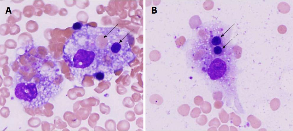
Figure 2 Hemophagocytosis in bone marrow.
A: High-power photomicrograph of Diff Quick stain of bone marrow smear showing two large macrophages with foamy cytoplasm within a background of mature erythrocytes. The macrophage on the right has phagocytosed a mature erythrocyte (pink cell with no nucleus, higher arrow) and a nucleated erythrocyte precursor (pink cell with deeply purple nucleus, lower arrow; B: high-power photomicrograph of another large macrophage with foamy cytoplasm containing two phagocytosed nucleated erythrocyte precursor cells (purple nucleus, two arrows).
- Citation: Cappell MS, Hader I, Amin M. Acute liver failure secondary to severe systemic disease from fatal hemophagocytic lymphohistiocytosis: Case report and systematic literature review. World J Hepatol 2018; 10(9): 629-636
- URL: https://www.wjgnet.com/1948-5182/full/v10/i9/629.htm
- DOI: https://dx.doi.org/10.4254/wjh.v10.i9.629


