Copyright
©The Author(s) 2018.
World J Hepatol. Jul 27, 2018; 10(7): 523-529
Published online Jul 27, 2018. doi: 10.4254/wjh.v10.i7.523
Published online Jul 27, 2018. doi: 10.4254/wjh.v10.i7.523
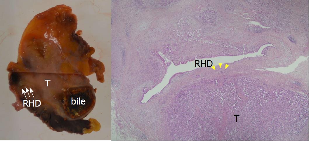
Figure 8 Pathological examination.
Pathological examination shows well-differentiated tubular adenocarcinoma of the gallbladder (T) with direct invasion of the liver parenchyma and the right hepatic duct (RHD: white arrow). The tumor cell invaded to the RHD wall, but not into the lumen (yellow arrowhead) (hematoxylin eosin saffron, original magnification × 20).
- Citation: Goto T, Terajima H, Yamamoto T, Uchida Y. Hepatectomy for gallbladder-cancer with unclassified anomaly of right-sided ligamentum teres: A case report and review of the literature. World J Hepatol 2018; 10(7): 523-529
- URL: https://www.wjgnet.com/1948-5182/full/v10/i7/523.htm
- DOI: https://dx.doi.org/10.4254/wjh.v10.i7.523


