Copyright
©The Author(s) 2018.
World J Hepatol. Jul 27, 2018; 10(7): 523-529
Published online Jul 27, 2018. doi: 10.4254/wjh.v10.i7.523
Published online Jul 27, 2018. doi: 10.4254/wjh.v10.i7.523
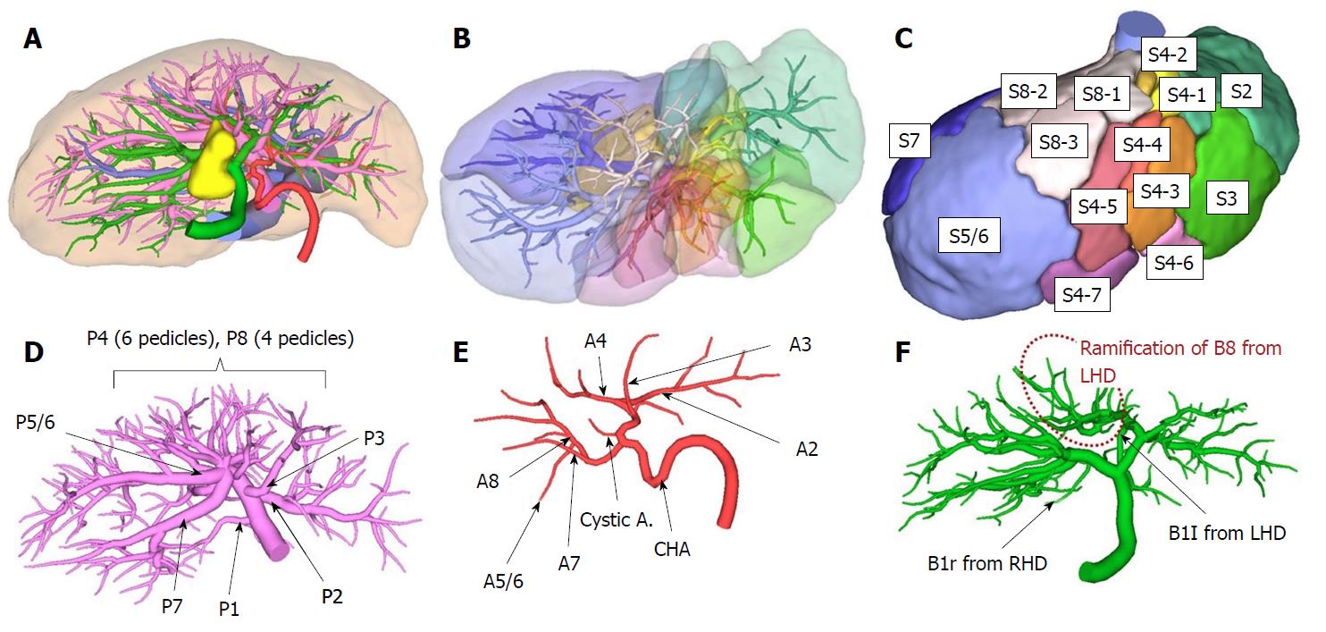
Figure 2 Preoperative simulation.
A: An “all-in-one” simulation image. The intrahepatic vasculature was reconstructed at the 4th order division level (red: hepatic artery, pink: portal vein, green: bile duct, yellow: Gallbladder, blue: hepatic vein); B and C: Simulated segmentation based on the portal venous flow. Couinaud’s definition was referred to for the naming of each segment; D: All segmental portal branches were ramified from the portal trunk. Interestingly, a common trunk of P5 and P6 was present; E: Hepatic arterial ramification without anomalous anatomy; F: The bile duct of segment 8 (B8) was ramified from the left hepatic duct, not from the right hepatic duct. RHD: Right hepatic duct; LHD: Left hepatic duct.
- Citation: Goto T, Terajima H, Yamamoto T, Uchida Y. Hepatectomy for gallbladder-cancer with unclassified anomaly of right-sided ligamentum teres: A case report and review of the literature. World J Hepatol 2018; 10(7): 523-529
- URL: https://www.wjgnet.com/1948-5182/full/v10/i7/523.htm
- DOI: https://dx.doi.org/10.4254/wjh.v10.i7.523


