Copyright
©The Author(s) 2018.
World J Hepatol. Jul 27, 2018; 10(7): 523-529
Published online Jul 27, 2018. doi: 10.4254/wjh.v10.i7.523
Published online Jul 27, 2018. doi: 10.4254/wjh.v10.i7.523
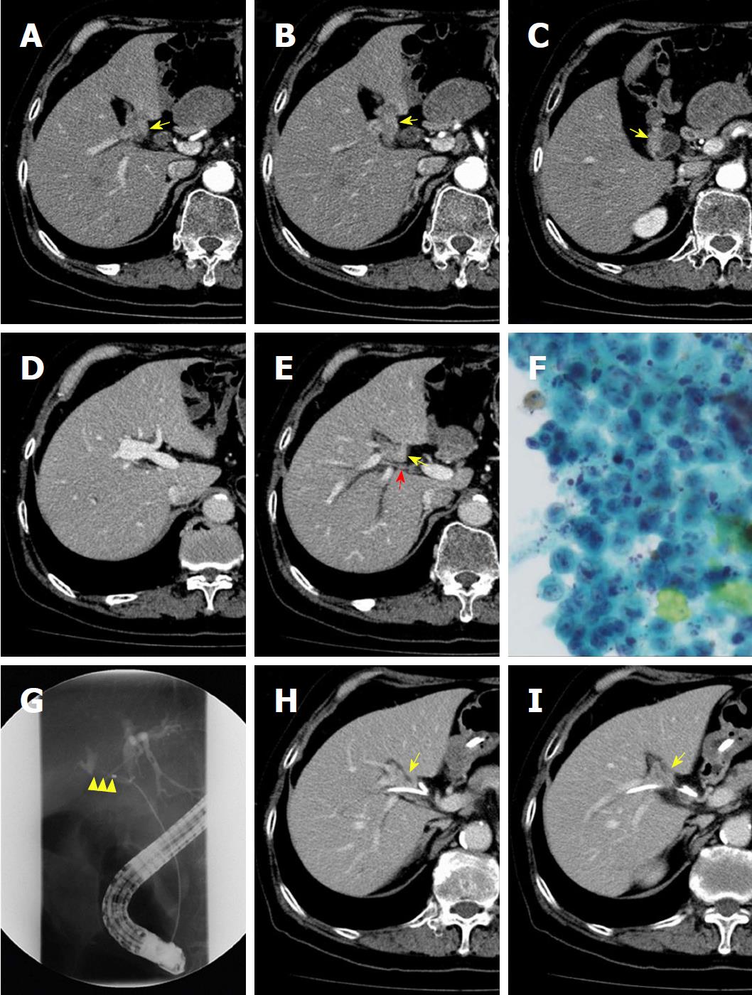
Figure 1 Contrast-enhanced computed tomography, cytological examination of bile, and endoscopic retrograde cholangiography.
A, B and C: The highly atrophied gallbladder (yellow arrow) had tumor-like localized wall thickness enhanced by contrast medium; D: The cul-de-sac of the right-sided umbilical portion is shown; E: The gallbladder (yellow arrow) is located on the left-sided liver bed. Direct infiltration of the tumor into the right hepatic artery (red arrow) is not evident; F: Cytological examination of bile obtained from endoscopic retrograde gallbladder drainage demonstrated atypical cells with a high nucleo-cytoplasmic ratio, strongly suggesting adenocarcinoma; G: Endoscopic retrograde cholangiography shows severe stenosis of the right hepatic duct (yellow arrowhead); H and I: The gallbladder tumor (yellow arrow) directly spreads to the nearby right hepatic duct where an endoscopic nasobiliary tube was placed.
- Citation: Goto T, Terajima H, Yamamoto T, Uchida Y. Hepatectomy for gallbladder-cancer with unclassified anomaly of right-sided ligamentum teres: A case report and review of the literature. World J Hepatol 2018; 10(7): 523-529
- URL: https://www.wjgnet.com/1948-5182/full/v10/i7/523.htm
- DOI: https://dx.doi.org/10.4254/wjh.v10.i7.523


