Copyright
©The Author(s) 2018.
World J Hepatol. Jul 27, 2018; 10(7): 485-495
Published online Jul 27, 2018. doi: 10.4254/wjh.v10.i7.485
Published online Jul 27, 2018. doi: 10.4254/wjh.v10.i7.485
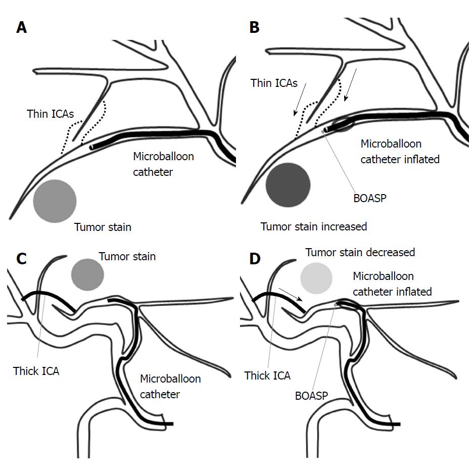
Figure 4 A schematic illustration of the balloon-occluded transcatheter arterial chemoembolization procedure.
A: A microballoon catheter is inserted into the tumor-feeding artery and inflated to occlude it. If the distal arterial blood flow cannot be stopped, the intrahepatic collateral arteries (ICAs) may be maintaining the distal arterial blood flow; B: When the ICAs are thin, the balloon-occluded arterial stump pressure (BOASP) is remarkably reduced compared to the arterial pressure without balloon occlusion. In this situation, we might observe increased tumor enhancement on selective computed tomography during hepatic angiography (CTHA) after balloon occlusion. A remarkable reduction in the BOASP is thought to allow us to inject the lipiodol emulsion (LE) under higher pressure, leading to the denser LE accumulation in targeted tumors. The BOASP after the injection of LE and gelatin particles is significantly higher than that before treatment; C and D: When the ICAs are thick, the BOASP is expected to be mildly reduced or unchanged because of the adequate blood flow from a thick ICA. In this situation, we might observe decreased tumor enhancement on CTHA after balloon occlusion. We would probably inject the LE under lower pressure in such a situation, thereby achieving a less-dense LE accumulation.
- Citation: Hatanaka T, Arai H, Kakizaki S. Balloon-occluded transcatheter arterial chemoembolization for hepatocellular carcinoma. World J Hepatol 2018; 10(7): 485-495
- URL: https://www.wjgnet.com/1948-5182/full/v10/i7/485.htm
- DOI: https://dx.doi.org/10.4254/wjh.v10.i7.485


