Copyright
©The Author(s) 2018.
World J Hepatol. Jul 27, 2018; 10(7): 462-473
Published online Jul 27, 2018. doi: 10.4254/wjh.v10.i7.462
Published online Jul 27, 2018. doi: 10.4254/wjh.v10.i7.462
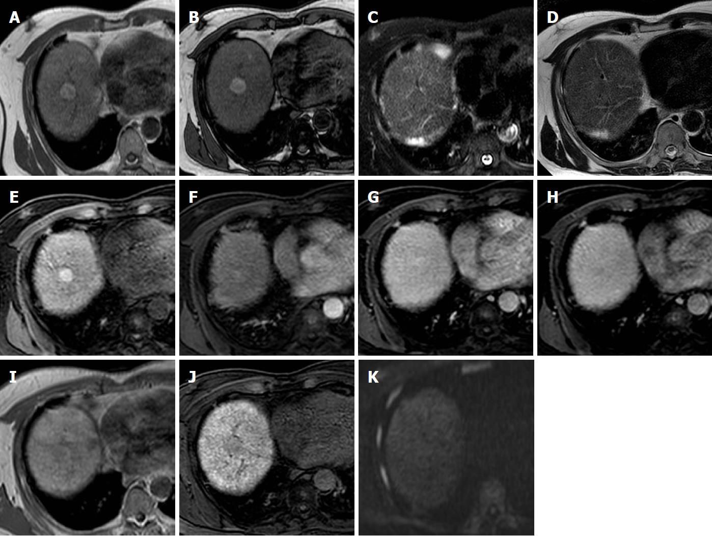
Figure 3 High-grade dysplastic nodule.
Gd-EOB-DTPA enhanced MR images of a 57 years old cirrhotic patient with a liver nodule in the VIII segment. A and B: Axial T1-weighted sequences both ”in phase” and “out of phase” show a hyperintense nodule; C and D: On T2-weighted image with and without fat saturation the nodule appears as isointense; E-H: during the dynamic contrast-enhanced images the nodule shows a slight enhancement in the arterial phase, without wash-out in portal and delayed phases; I and J: Diffusion weighted image demonstrate no restriction to the diffusion; K: In hepatobiliary phase the nodule is hypointense in comparison to the surrounding liver parenchyma. The MRI features, suggestive of high grade dysplastic nodule, have been later confirmed by the histological examination. MRI: Magnetic resonance imaging.
- Citation: Inchingolo R, Faletti R, Grazioli L, Tricarico E, Gatti M, Pecorelli A, Ippolito D. MR with Gd-EOB-DTPA in assessment of liver nodules in cirrhotic patients. World J Hepatol 2018; 10(7): 462-473
- URL: https://www.wjgnet.com/1948-5182/full/v10/i7/462.htm
- DOI: https://dx.doi.org/10.4254/wjh.v10.i7.462


