Copyright
©The Author(s) 2018.
World J Hepatol. Apr 27, 2018; 10(4): 402-408
Published online Apr 27, 2018. doi: 10.4254/wjh.v10.i4.402
Published online Apr 27, 2018. doi: 10.4254/wjh.v10.i4.402
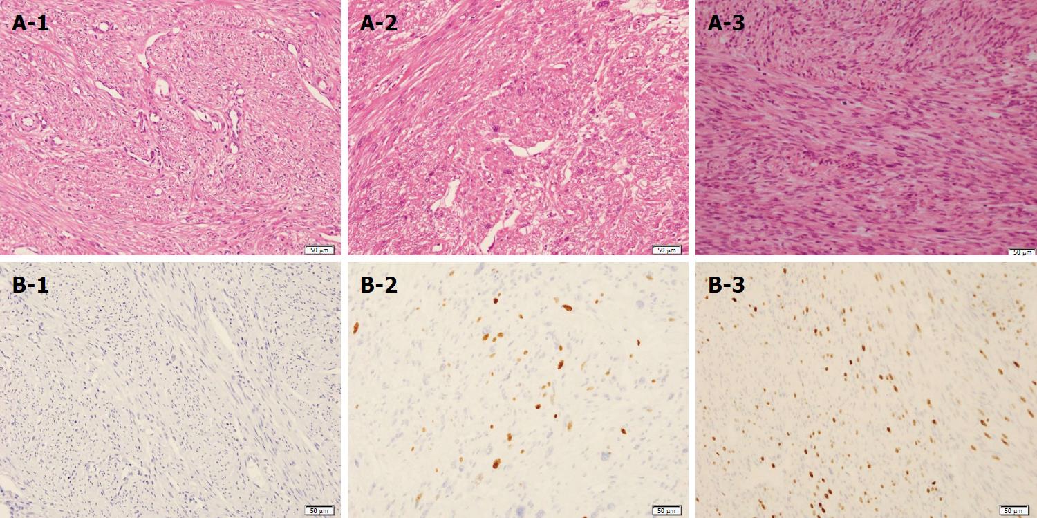
Figure 4 Pathological reconfirmation of uterine leiomyoma and broad ligament with STUMP and liver lesion with leiomyosarcoma.
MF was absent in an HE stain of the uterine leiomyoma (A-1). The number of MFs was 1-5 MFs / 10 HPFs in the broad ligament (A-2) and more than 10 MFs/10 HPFs in the liver mass (A-3). Ki-67-positive cells were not seen in the uterine leiomyoma (B-1). The MIB-1 index was approximately 5% of the broad ligament and approximately 35% of the liver mass (B-2) (B-3). The pathological findings of MFs and the MIB-1 index showed malignant transformation from STUMP to leiomyosarcoma. Scale bars: 50 μm.
- Citation: Fukui K, Takase N, Miyake T, Hisano K, Maeda E, Nishimura T, Abe K, Kozuki A, Tanaka T, Harada N, Takamatsu M, Kaneda K. Review of the literature laparoscopic surgery for metastatic hepatic leiomyosarcoma associated with smooth muscle tumor of uncertain malignant potential: Case report. World J Hepatol 2018; 10(4): 402-408
- URL: https://www.wjgnet.com/1948-5182/full/v10/i4/402.htm
- DOI: https://dx.doi.org/10.4254/wjh.v10.i4.402


