Copyright
©The Author(s) 2018.
World J Hepatol. Apr 27, 2018; 10(4): 402-408
Published online Apr 27, 2018. doi: 10.4254/wjh.v10.i4.402
Published online Apr 27, 2018. doi: 10.4254/wjh.v10.i4.402
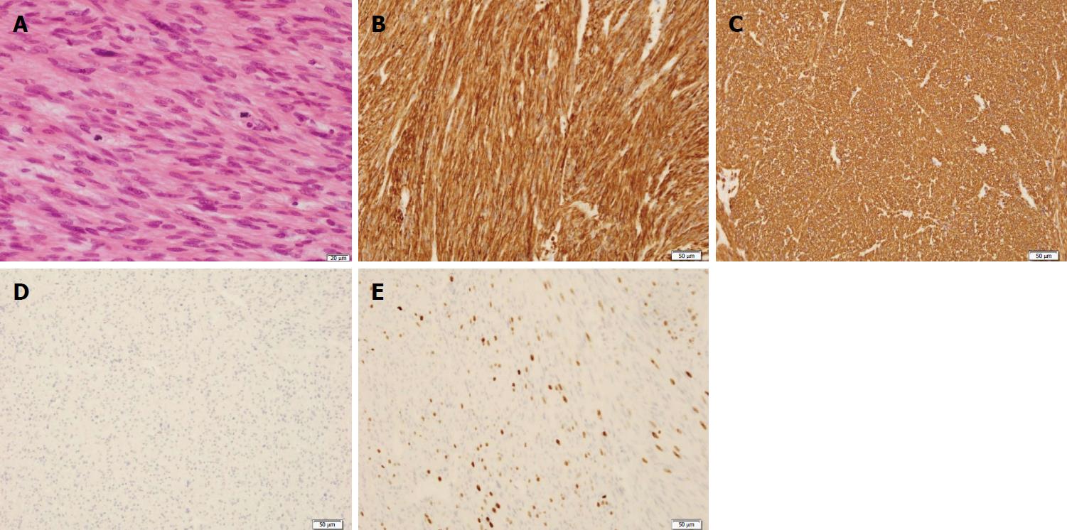
Figure 3 Pathological findings of the resected specimen.
A: HE stain showed the proliferation of spindle-shaped cells with ≥ 10 MFs-/-10 HPFs and diffuse moderate-to-severe atypia without coagulative tumor cell necrosis; B: Immunohistochemical findings were strongly positive for SMA; C: Immunohistochemical findings were strongly positive for desmin; D: Immunohistochemical findings were strongly positive for negative for c-kit; E: Approximately 35% ki-67 positive cells were seen in the lesion. Scale bars: 20 μm (A), 50 μm (B-E).
- Citation: Fukui K, Takase N, Miyake T, Hisano K, Maeda E, Nishimura T, Abe K, Kozuki A, Tanaka T, Harada N, Takamatsu M, Kaneda K. Review of the literature laparoscopic surgery for metastatic hepatic leiomyosarcoma associated with smooth muscle tumor of uncertain malignant potential: Case report. World J Hepatol 2018; 10(4): 402-408
- URL: https://www.wjgnet.com/1948-5182/full/v10/i4/402.htm
- DOI: https://dx.doi.org/10.4254/wjh.v10.i4.402


