Copyright
©The Author(s) 2018.
World J Hepatol. Apr 27, 2018; 10(4): 402-408
Published online Apr 27, 2018. doi: 10.4254/wjh.v10.i4.402
Published online Apr 27, 2018. doi: 10.4254/wjh.v10.i4.402
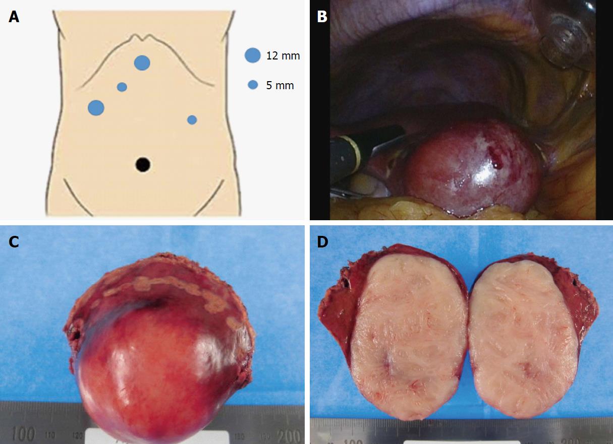
Figure 2 Ultrasound-guided pure laparoscopic partial liver resection (Segment 4) and surgical specimen.
A: Five ports were placed for liver partial resection. Three ports were placed around the epigastric region as working ports (blue circles). An umbilical port was used as the camera port (black circle). The resected partial liver was retrieved from the umbilical port with auxiliary incision. A Pringle maneuver was used for the left lateral port; B: We performed ultrasound-guided pure laparoscopic partial liver resection with vessel-sealing devices (LigaSure™ Maryland Jaw 37 cm Laparoscopic Sealer/Divider, Medtronic, Dublin, Ireland) using the crush clamping method and Pringle maneuver; C: Macroscopic image of the resected specimen showed a smooth surface mass; D: Cross-section of the resected specimen showed a milky-white-colored solid mass without necrotic lesion.
- Citation: Fukui K, Takase N, Miyake T, Hisano K, Maeda E, Nishimura T, Abe K, Kozuki A, Tanaka T, Harada N, Takamatsu M, Kaneda K. Review of the literature laparoscopic surgery for metastatic hepatic leiomyosarcoma associated with smooth muscle tumor of uncertain malignant potential: Case report. World J Hepatol 2018; 10(4): 402-408
- URL: https://www.wjgnet.com/1948-5182/full/v10/i4/402.htm
- DOI: https://dx.doi.org/10.4254/wjh.v10.i4.402


