Copyright
©The Author(s) 2018.
World J Hepatol. Apr 27, 2018; 10(4): 402-408
Published online Apr 27, 2018. doi: 10.4254/wjh.v10.i4.402
Published online Apr 27, 2018. doi: 10.4254/wjh.v10.i4.402
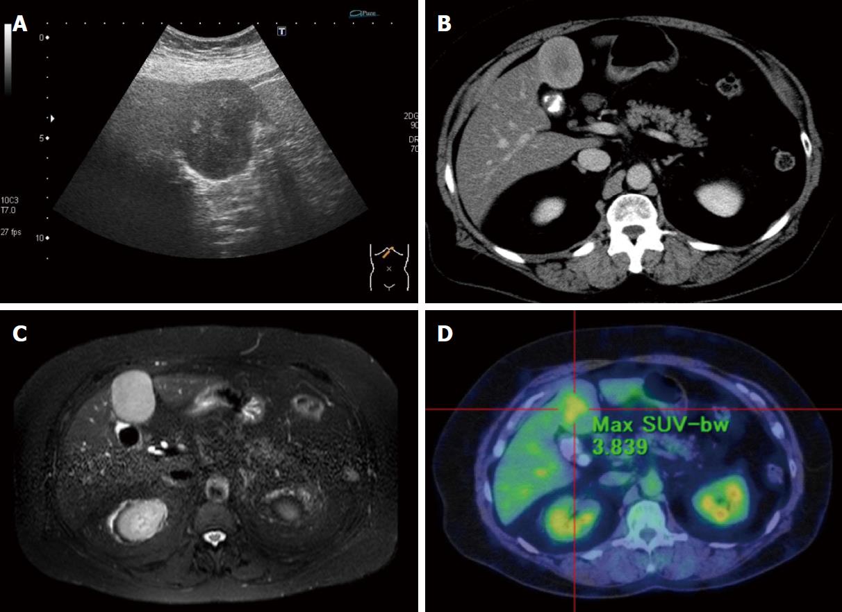
Figure 1 Evaluation of clinical findings.
A: Ultrasonography of the liver showed a very-low-echoic smooth mass (60 mm × 40 mm) with a heterogeneous high-echoic region in the median section of the left lobe of the liver (Segment 4); B: Axial delayed phase CE-CT showed the lesion to be gradually enhanced heterogeneously; C: The lesion showed a hyperintense heterogeneous region on axial T2-weighted EOB-MRI; D: The lesion showed obvious metabolically active foci by 18-fluorodeoxyglucose-PET-CT evaluation, while other lesions were not detected.
- Citation: Fukui K, Takase N, Miyake T, Hisano K, Maeda E, Nishimura T, Abe K, Kozuki A, Tanaka T, Harada N, Takamatsu M, Kaneda K. Review of the literature laparoscopic surgery for metastatic hepatic leiomyosarcoma associated with smooth muscle tumor of uncertain malignant potential: Case report. World J Hepatol 2018; 10(4): 402-408
- URL: https://www.wjgnet.com/1948-5182/full/v10/i4/402.htm
- DOI: https://dx.doi.org/10.4254/wjh.v10.i4.402


