Copyright
©The Author(s) 2018.
World J Hepatol. Feb 27, 2018; 10(2): 213-221
Published online Feb 27, 2018. doi: 10.4254/wjh.v10.i2.213
Published online Feb 27, 2018. doi: 10.4254/wjh.v10.i2.213
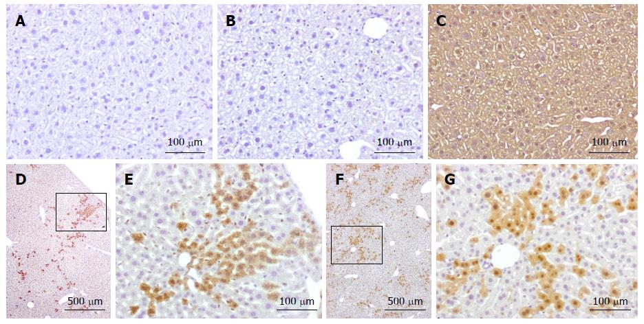
Figure 2 Immunohistochemical analysis of tdTomato expression in livers of chimeras and controls.
For tdTomato detection, 3 μm slices were stained with anti-RFP antibody (ab124754: Abcam, Cambridge, United Kingdom) diluted 1:100 in PBS containing 0.05% Tween 20. Detection was performed using Mach 1 HRP-polymer (Biocare Medical, Concord, CA, United States) incubation followed by the revelation with Betazoid DAB (Biocare Medical). A: C57BL/6J wild-type mouse; B: Inactive tdTomato mouse; C: Cre+/tdTomato+ double transgenic mouse; D-G: Cre:tdTomato chimera 1 and 2, respectively. Wild type and Inactive Tomato mice are completely negative; Cre+/tdTomato+ are completely positive due to activation of the tdTomato gene in all cells. In the chimeras, many cells are negative, but a fraction shows clear expression of the tdTomato gene, which has been activated by the co-expression of Cre in fused cells. E and G: higher magnification views of the boxed area in D and F respectively.
- Citation: Lizier M, Castelli A, Montagna C, Lucchini F, Vezzoni P, Faggioli F. Cell fusion in the liver, revisited. World J Hepatol 2018; 10(2): 213-221
- URL: https://www.wjgnet.com/1948-5182/full/v10/i2/213.htm
- DOI: https://dx.doi.org/10.4254/wjh.v10.i2.213


