Copyright
©The Author(s) 2018.
World J Hepatol. Jan 27, 2018; 10(1): 166-171
Published online Jan 27, 2018. doi: 10.4254/wjh.v10.i1.166
Published online Jan 27, 2018. doi: 10.4254/wjh.v10.i1.166
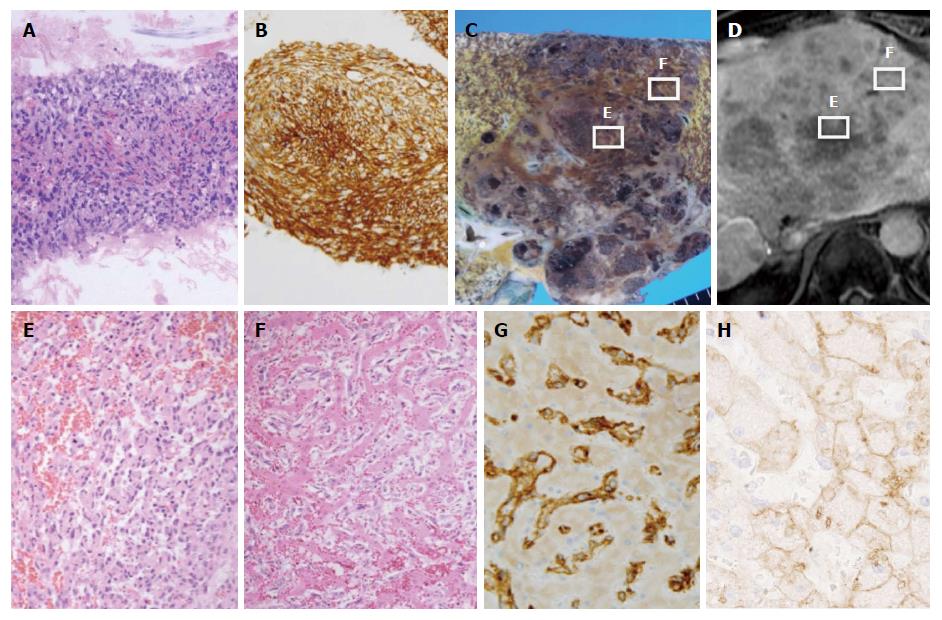
Figure 2 Histological findings from a liver tumor biopsy revealed the solid proliferation of spindle cells.
A: Hematoxylin and eosin (HE) staining, × 20; B: CD34 staining, × 20; C: Macroscopically, a large tumor and many satellite nodules were observed in the liver; D: A hepatobiliary phase image obtained using gadolinium-ethoxybenzyl-diethylenetriamine-enhanced magnetic resonance imaging, depict almost the same plane of C; E: The proliferation of atypical spindle cells and hemorrhage were seen (HE staining, × 100); F: Tumor cells had spread to the sinusoids in most of the liver, although normal hepatic cells remained (HE staining, × 100); G: Tumor cells were positive for CD34 (CD34 staining, × 400); H: Tumor cells were negative for OATP1B3, and hepatic cells were positive for OATP1B3 (OATP1B3 staining, × 400).
- Citation: Hayashi M, Kawana S, Sekino H, Abe K, Matsuoka N, Kashiwagi M, Okai K, Kanno Y, Takahashi A, Ito H, Hashimoto Y, Ohira H. Contrast uptake in primary hepatic angiosarcoma on gadolinium-ethoxybenzyl-diethylenetriamine pentaacetic acid-enhanced magnetic resonance imaging in the hepatobiliary phase. World J Hepatol 2018; 10(1): 166-171
- URL: https://www.wjgnet.com/1948-5182/full/v10/i1/166.htm
- DOI: https://dx.doi.org/10.4254/wjh.v10.i1.166


