Copyright
©2009 Baishideng.
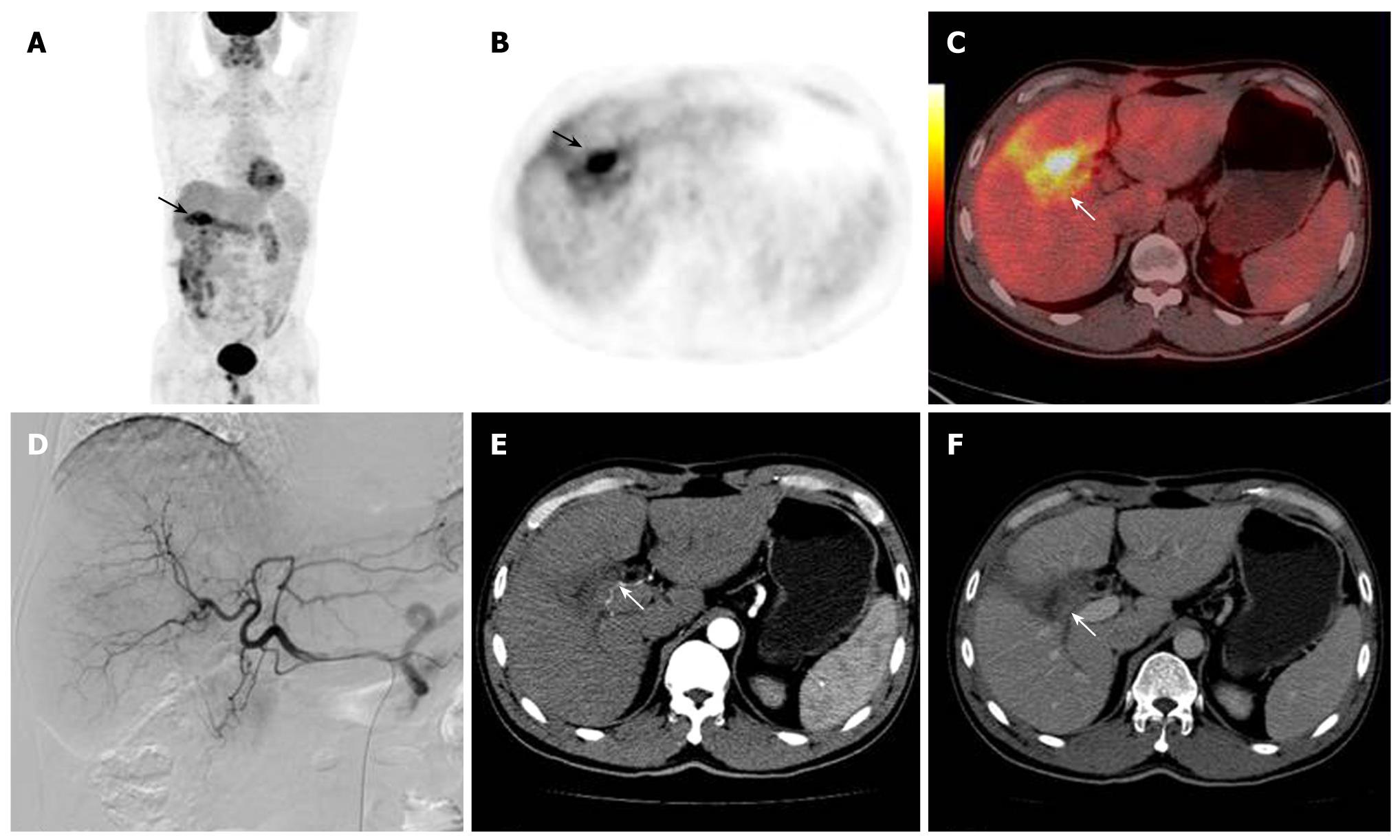
Figure 4 A 48-year man who had HCC resection 2 mo before the imaging, followed by TACE 40 d later.
PET and PET/CT fused images revealed highly metabolic lesions (arrows, A-C). Both contrast-enhanced axial CT imaging and arteriography failed to show lesions in the right lobe of the liver (arrows, D-F). These findings of PET/CT were later verified as inflammatory by post-operative tissue examination.
- Citation: Sun L, Guan YS, Pan WM, Luo ZM, Wei JH, Zhao L, Wu H. Metabolic restaging of hepatocellular carcinoma using whole-body 18F-FDG PET/CT. World J Hepatol 2009; 1(1): 90-97
- URL: https://www.wjgnet.com/1948-5182/full/v1/i1/90.htm
- DOI: https://dx.doi.org/10.4254/wjh.v1.i1.90


