Copyright
©2009 Baishideng.
World J Hepatol. Oct 31, 2009; 1(1): 103-109
Published online Oct 31, 2009. doi: 10.4254/wjh.v1.i1.103
Published online Oct 31, 2009. doi: 10.4254/wjh.v1.i1.103
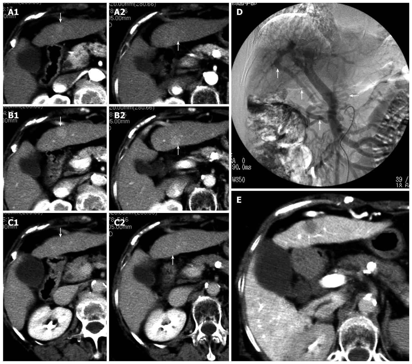
Figure 1 Dynamic computed tomography findings at the onset of double cancer with hepatocellular and cholangiocellular carcinomas.
Two tumors, ~1 cm in diameter, were detected on the ventral and dorsal sides of S3. Arrows indicate the tumors (A1, A2 arterial phase; B1, B2 parenchymal phase; C1, C2 delayed phase). Angiographic findings at the onset of double cancer; D: A superior mesenteric arterial angiogram showing a large shunt through the epigastric vein during the portal phase (the arrows); E: Computed tomography during arterial portography (CTAP) showing a ~1 cm defect on the ventral side of S3 (the arrow), which was diagnosed as a classic hepatocellular carcinoma. No defect can be seen on the dorsal side of S3.
- Citation: Watanabe T, Sakata J, Ishikawa T, Shirai Y, Suda T, Hirono H, Hasegawa K, Soga K, Shibasaki K, Saito Y, Umezu H. Synchronous development of HCC and CCC in the same subsegment of the liver in a patient with type C liver cirrhosis. World J Hepatol 2009; 1(1): 103-109
- URL: https://www.wjgnet.com/1948-5182/full/v1/i1/103.htm
- DOI: https://dx.doi.org/10.4254/wjh.v1.i1.103


