Copyright
©The Author(s) 2015.
World J Hepatol. Jun 28, 2015; 7(12): 1694-1700
Published online Jun 28, 2015. doi: 10.4254/wjh.v7.i12.1694
Published online Jun 28, 2015. doi: 10.4254/wjh.v7.i12.1694
Figure 1 Contrast enhanced computed tomography in patient with multinodular hepatocellular carcinoma before the degradable starch microspheres transcatheter arterial chemoembolization not in agreement with new-Milan-criteria.
The enhanced computed tomography images show 5 hypervascular lesions, in the patients with a score of 8 using the new-Milan-criteria. A: Hepatocellular carcinoma (HCC) on VII segment with maximum diameter of 14 mm1; B: HCC on VIII segment with maximum diameter of 10 mm1; C: HCC on VII segment with maximum diameter of 12 mm1; D: HCC on VIII segment with maximum diameter of 31 mm1; E: HCC on IV-II segment with maximum diameter of 14 mm.
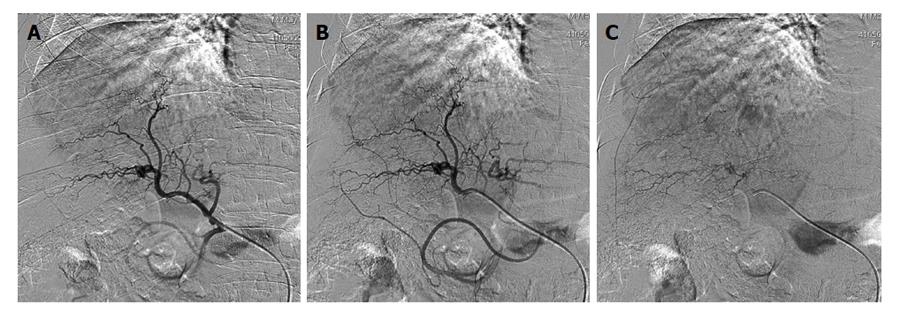
Figure 2 Hepatic intraprocedural angiograms show staining of multiple tumors, which are diagnosed as hepatocellular carcinoma.
A and B: Early arterial digital subtraction angiography (DSA) phase; C: Late parenchymal DSA phase.
Figure 3 Enhanced computed tomography control after degradable starch microspheres transcatheter arterial chemoembolization shows a complete response of 4/5 lesions in well downstaged patient.
After degradable starch microspheres transcatheter arterial chemoembolization patients had a new-Milan-criteria score of 4. A: Complete response of hepatocellular carcinoma (HCC) nodule on VIII segment; B: On VII segment (white arrows); C and D: Partial response of HCC nodules (red and white arrows).
- Citation: Orlacchio A, Chegai F, Merolla S, Francioso S, Giudice CD, Angelico M, Tisone G, Simonetti G. Downstaging disease in patients with hepatocellular carcinoma outside up-to-seven criteria: Strategies using degradable starch microspheres transcatheter arterial chemo-embolization. World J Hepatol 2015; 7(12): 1694-1700
- URL: https://www.wjgnet.com/1948-5182/full/v7/i12/1694.htm
- DOI: https://dx.doi.org/10.4254/wjh.v7.i12.1694


