Copyright
©The Author(s) 2022.
World J Hepatol. May 27, 2022; 14(5): 944-955
Published online May 27, 2022. doi: 10.4254/wjh.v14.i5.944
Published online May 27, 2022. doi: 10.4254/wjh.v14.i5.944
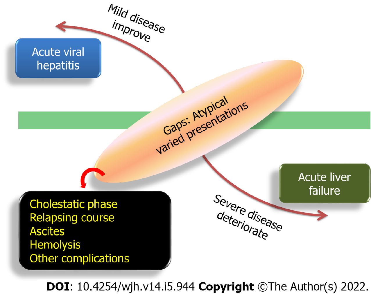
Figure 1 Spectrum of acute viral hepatitis.
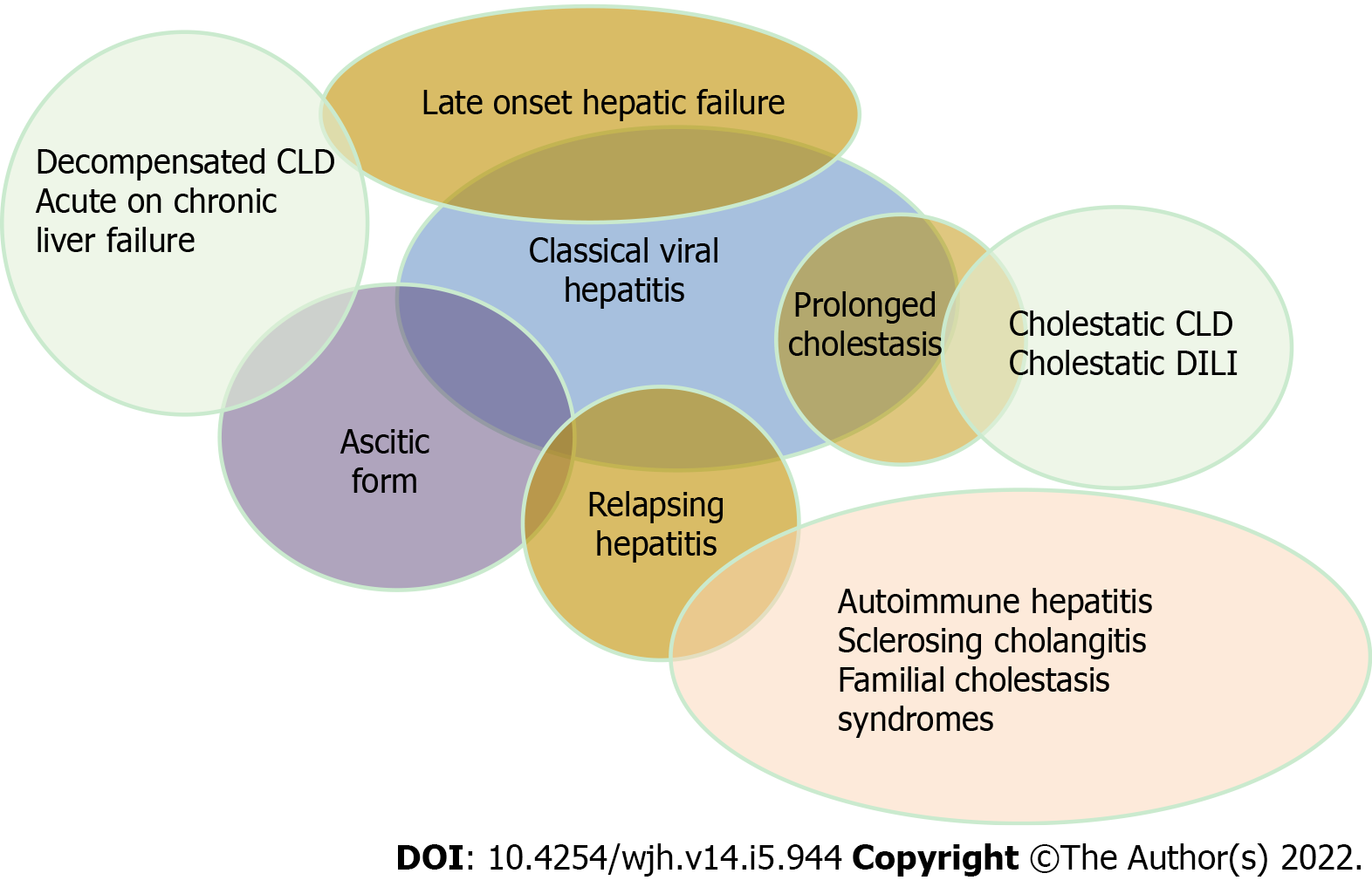
Figure 2 Overlapping atypical manifestations of acute viral hepatitis.
CLD: Chronic liver disease; DILI: Drug-induced liver injury.
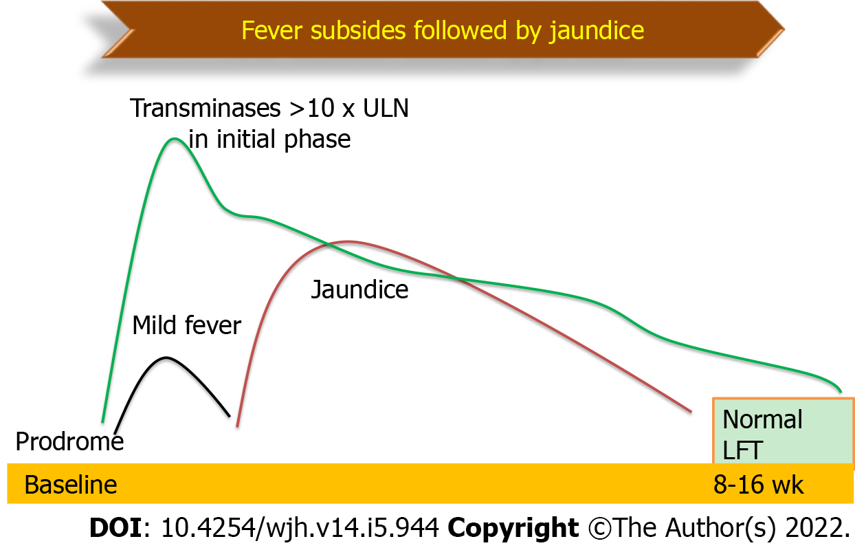
Figure 3 Natural history of classical acute viral hepatitis.
LFT: Liver function tests.
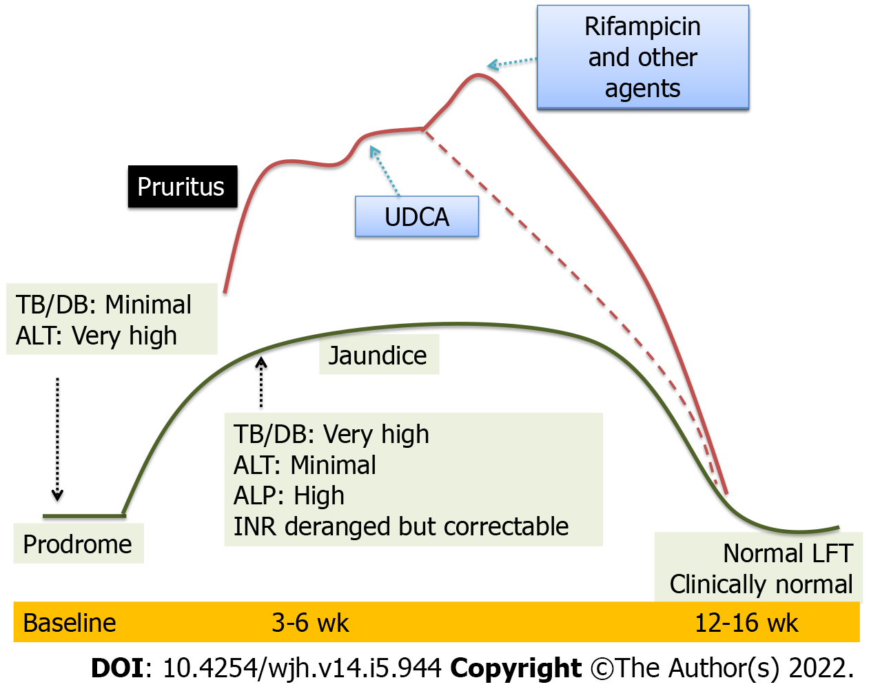
Figure 4 Natural history of prolonged cholestasis in acute viral hepatitis.
LFT: Liver function tests; UDCA: Ursodeoxycholic acid; ALT: Alanine aminotransferase; TB/DB: Tuberculosis/Disulfide bond; ALP: Alkaline phosphatase; INR: International normalized ratio.
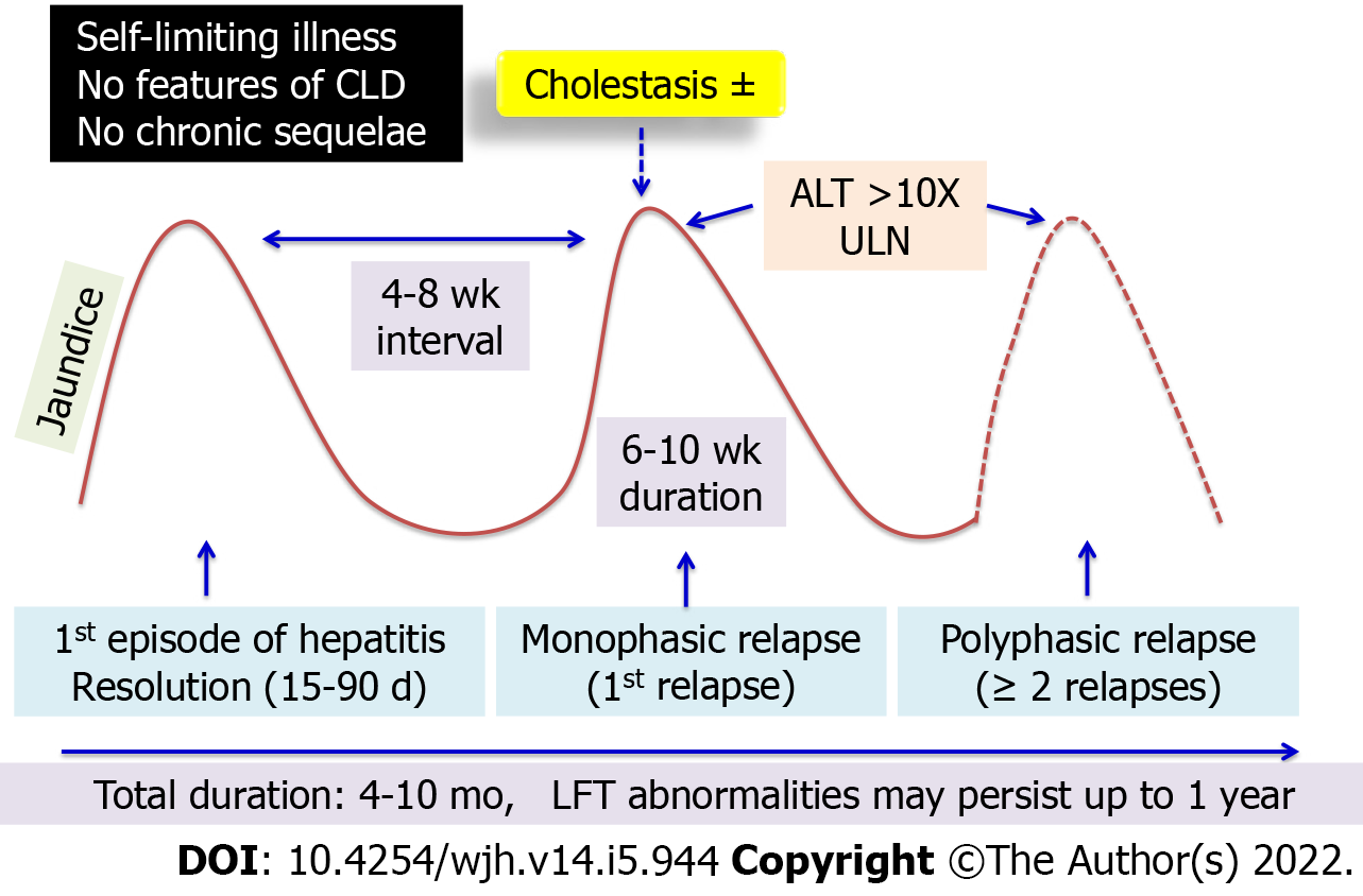
Figure 5 Natural history of relapsing hepatitis in acute viral hepatitis.
CLD: Chronic liver disease; ALT: Alanine aminotransferase; LFT: Liver function tests.
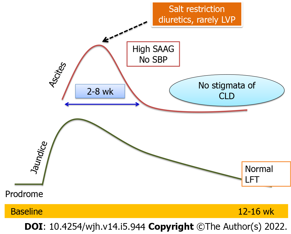
Figure 6 Natural history of ascites in acute viral hepatitis.
LVP: Levator veli palatine; SAAG: Serum-ascites albumin gradient; SBP: Systolic blood pressure; CLD: Chronic liver disease; LFT: Liver function tests.
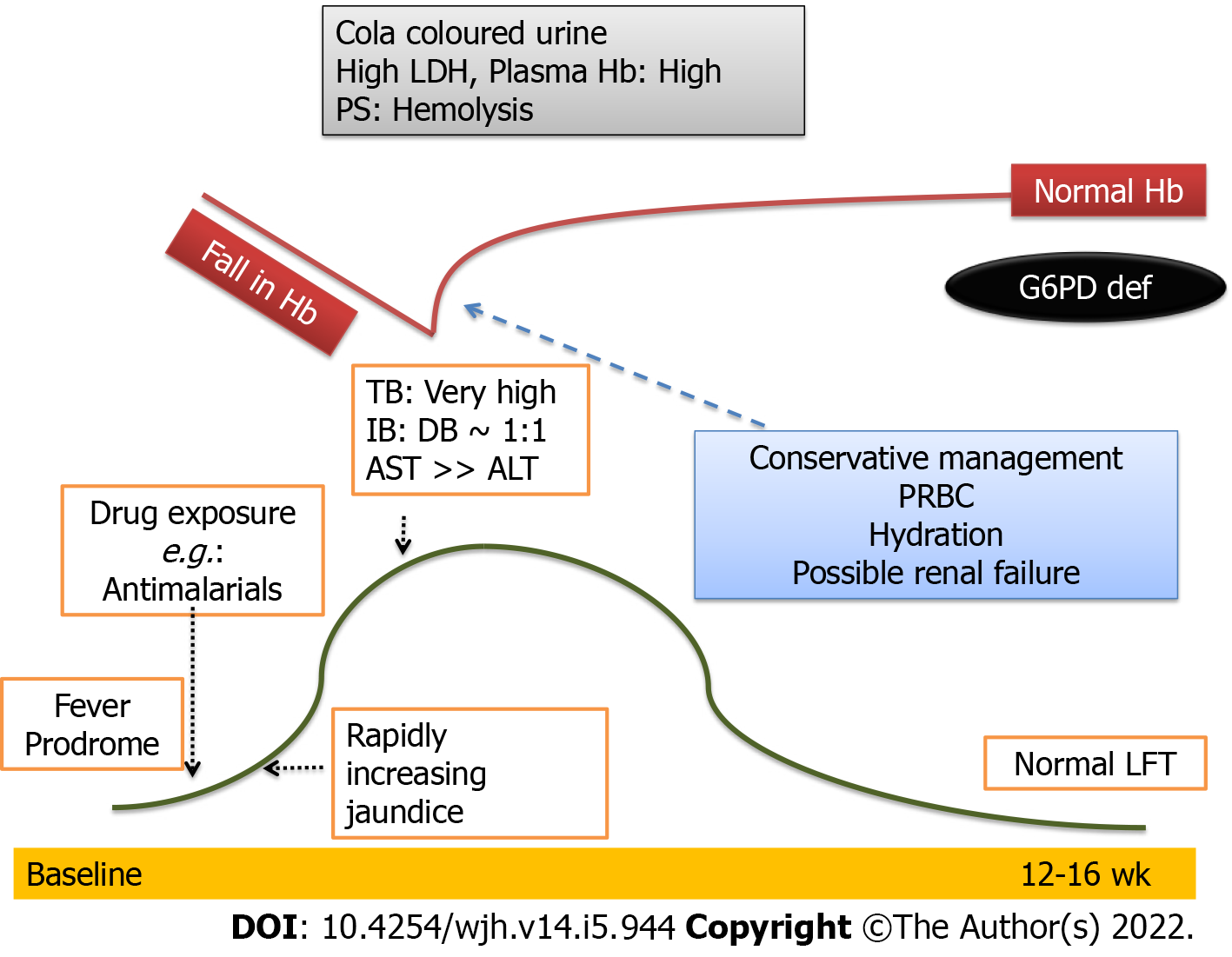
Figure 7 Natural history of intravascular hemolysis (G6PD deficiency) in acute viral hepatitis.
LDH: Lactate dehydrogenase; TB: Tuberculosis; IB: Illipe butter; DB: Disulfide bond; AST: Aspartate aminotransferase; ALT: Alanine aminotransferase; PRBC: Packed red blood cells; LFT: Liver function tests.
- Citation: Sarma MS, Ravindranath A. Pediatric acute viral hepatitis with atypical variants: Clinical dilemmas and natural history. World J Hepatol 2022; 14(5): 944-955
- URL: https://www.wjgnet.com/1948-5182/full/v14/i5/944.htm
- DOI: https://dx.doi.org/10.4254/wjh.v14.i5.944


