Copyright
©The Author(s) 2017.
World J Stem Cells. Aug 26, 2017; 9(8): 127-132
Published online Aug 26, 2017. doi: 10.4252/wjsc.v9.i8.127
Published online Aug 26, 2017. doi: 10.4252/wjsc.v9.i8.127
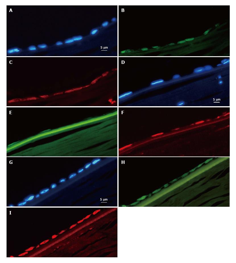
Figure 2 Endothelial parts of three corneal sections: A-C, D-F, and G-I.
A: Nuclei through a 4‘,6-diamidino-2-phenylindole (DAPI) filter; B: The same cells through a fluorescein isothiocyanate (FITC) filter [green fluorescent protein (GFP) fluorescence]; C: Expression of the tight junction protein Zona Occludens protein 1 (ZO-1) [tetramethylrhodamine (TRITC) filter]; D: Nuclei seen through a DAPI filter; E: GFP expression (using an FITC filter); F: Expression of sodium potassium ATPase (NaKATPase) (using a TRITC filter); G: Nuclei seen through a DAPI filter; H: GFP expression (FITC filter); I: Expression of the cornea-related protein paired box 6 (PAX-6).
- Citation: Hanson C, Arnarsson A, Hardarson T, Lindgård A, Daneshvarnaeini M, Ellerström C, Bruun A, Stenevi U. Transplanting embryonic stem cells onto damaged human corneal endothelium. World J Stem Cells 2017; 9(8): 127-132
- URL: https://www.wjgnet.com/1948-0210/full/v9/i8/127.htm
- DOI: https://dx.doi.org/10.4252/wjsc.v9.i8.127


