Copyright
©The Author(s) 2017.
World J Stem Cells. Jul 26, 2017; 9(7): 98-106
Published online Jul 26, 2017. doi: 10.4252/wjsc.v9.i7.98
Published online Jul 26, 2017. doi: 10.4252/wjsc.v9.i7.98
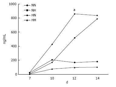
Figure 1 Osteocalcin secretion.
In cells exposed to hypoxia (5% O2) for 7 d followed by normoxia (21% O2) for 7 d (HN), the level of osteocalcin increased rapidly from day 10, peaking at day 12, as compared with cells in the other groups. NN, normoxia for 14 d; NH, normoxia for 7 d followed by hypoxia for 7 d; and HH, hypoxia for 14 d. aP < 0.05.
- Citation: Inagaki Y, Akahane M, Shimizu T, Inoue K, Egawa T, Kira T, Ogawa M, Kawate K, Tanaka Y. Modifying oxygen tension affects bone marrow stromal cell osteogenesis for regenerative medicine. World J Stem Cells 2017; 9(7): 98-106
- URL: https://www.wjgnet.com/1948-0210/full/v9/i7/98.htm
- DOI: https://dx.doi.org/10.4252/wjsc.v9.i7.98


