Copyright
©The Author(s) 2016.
World J Stem Cells. May 26, 2016; 8(5): 202-215
Published online May 26, 2016. doi: 10.4252/wjsc.v8.i5.202
Published online May 26, 2016. doi: 10.4252/wjsc.v8.i5.202
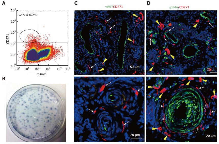
Figure 6 Specific markers for ovine e-mesenchymal stem cells.
Flow cytometry plot of ovine endometrial cells immunolabelled with CD271 and CD49f antibodies (A). The CD271+CD49f- population enriches; Clonogenic stromal cells (B); Immunofluorescence images of ovine endometrium stained with CD271 (red) and vascular markers reveals their in vivo perivascular location in the adventitia of veins and arteries (C, D); vWF an endothelial marker (green), showing CD271+ cells are perivascular but not pericytes (C); αSMA, a perivascular marker (green) showing CD271+ cells located adjacent to αSMA+ cells in the adventitia of vessels rather than expressing αSMA themselves (D). White arrows: perivascular CD271+ cells; yellow arrows: CD271+ cells not associated with vessels (reproduced from ref.[81] with permission). vWF: Von Willebrand factor; αSMA: Alpha smooth muscle actin.
- Citation: Emmerson SJ, Gargett CE. Endometrial mesenchymal stem cells as a cell based therapy for pelvic organ prolapse. World J Stem Cells 2016; 8(5): 202-215
- URL: https://www.wjgnet.com/1948-0210/full/v8/i5/202.htm
- DOI: https://dx.doi.org/10.4252/wjsc.v8.i5.202


