Copyright
©The Author(s) 2016.
World J Stem Cells. May 26, 2016; 8(5): 202-215
Published online May 26, 2016. doi: 10.4252/wjsc.v8.i5.202
Published online May 26, 2016. doi: 10.4252/wjsc.v8.i5.202
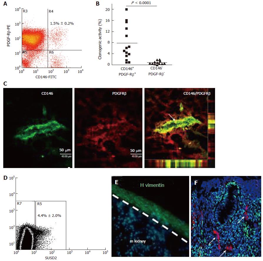
Figure 4 Specific enriching for endometrial mesenchymal stem cells.
Flow cytometry plot of CD146+PDGFRB+ fraction (A) which contains most of the clonogenic stromal cells (B) and reveals their pericyte identity in vivo (C); SUSD2+ cells in endometrial cell suspensions (D) which E reconstitute human vimentin+ stromal tissue when transplanted under the kidney capsule of NSG mice, and F have a perivascular location in human endometrium. SUSD2+ cells (red) do not express estrogen receptor-α (green), but endometrial stromal cells do (DNA blue). The white arrow indicates perivascular SUSD2+ cells (reproduced ref. [70,72,78] with permission).
- Citation: Emmerson SJ, Gargett CE. Endometrial mesenchymal stem cells as a cell based therapy for pelvic organ prolapse. World J Stem Cells 2016; 8(5): 202-215
- URL: https://www.wjgnet.com/1948-0210/full/v8/i5/202.htm
- DOI: https://dx.doi.org/10.4252/wjsc.v8.i5.202


