Copyright
©The Author(s) 2016.
World J Stem Cells. Feb 26, 2016; 8(2): 47-55
Published online Feb 26, 2016. doi: 10.4252/wjsc.v8.i2.47
Published online Feb 26, 2016. doi: 10.4252/wjsc.v8.i2.47
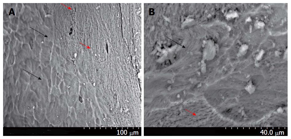
Figure 2 The attachment of platelet lysate-expanded bone marrow mesenchymal stem cells on orthoss scaffold.
BM MSCs at passage 2 were incubated with the scaffold by continuous mixing at 37 °C and 5% CO2 for 3 h with DMEM + 50 mL/L PL. The scaffold was then washed and incubated for 10 d with DMEM + 50 mL/L PL. SEM image illustrate MSCs on the scaffold (A). Higher magnification demonstrates MSC morphology on the scaffold surface (B). Black arrow: MSC cells; Red arrow: Orthoss scaffold. BM: Bone marrow; MSCs: Mesenchymal stem cells; PL: Platelet lysate.
- Citation: Altaie A, Owston H, Jones E. Use of platelet lysate for bone regeneration - are we ready for clinical translation? World J Stem Cells 2016; 8(2): 47-55
- URL: https://www.wjgnet.com/1948-0210/full/v8/i2/47.htm
- DOI: https://dx.doi.org/10.4252/wjsc.v8.i2.47


