Copyright
©The Author(s) 2016.
World J Stem Cells. Nov 26, 2016; 8(11): 384-395
Published online Nov 26, 2016. doi: 10.4252/wjsc.v8.i11.384
Published online Nov 26, 2016. doi: 10.4252/wjsc.v8.i11.384
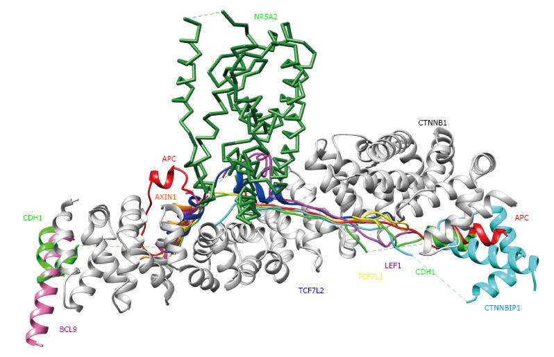
Figure 3 3D structures of β-catenin (CTNNB1) binding proteins.
Each CTNNB1 complex structure was superimposed onto the CTNNB1 structure in a complex with APC (PDB code: 1th1), using the program MATRAS (From Ref. [18]). Colors and PDB codes are summarized as follows: White: CTNNB1 (CTNB1_HUMAN, 1th1); red: APC (APC_HUMAN, 1th1); orange: AXIN1 (AXN_XELNA, 1qz7); hot pink: BCL9 (BCL9_HUMAN, 3sl9); green: CDH1 (CADH1_MOUSE. 1i7w); cyan: CTNNBIP1 (CNBP1_HUMAN, 1m1e); magenta: LEF1 (LEF1_MOUSE, 3oux); forest green: NR5A2 (NR5A2_HUMAN, 3tx7); yellow: TCF7L1 (T7L1A_XENLA, 1g3j); blue: TCF7L2 (TF7L2_HUMAN, 1jdh).
- Citation: Tanabe S, Kawabata T, Aoyagi K, Yokozaki H, Sasaki H. Gene expression and pathway analysis of CTNNB1 in cancer and stem cells. World J Stem Cells 2016; 8(11): 384-395
- URL: https://www.wjgnet.com/1948-0210/full/v8/i11/384.htm
- DOI: https://dx.doi.org/10.4252/wjsc.v8.i11.384


