Copyright
©The Author(s) 2016.
World J Stem Cells. Oct 26, 2016; 8(10): 355-366
Published online Oct 26, 2016. doi: 10.4252/wjsc.v8.i10.355
Published online Oct 26, 2016. doi: 10.4252/wjsc.v8.i10.355
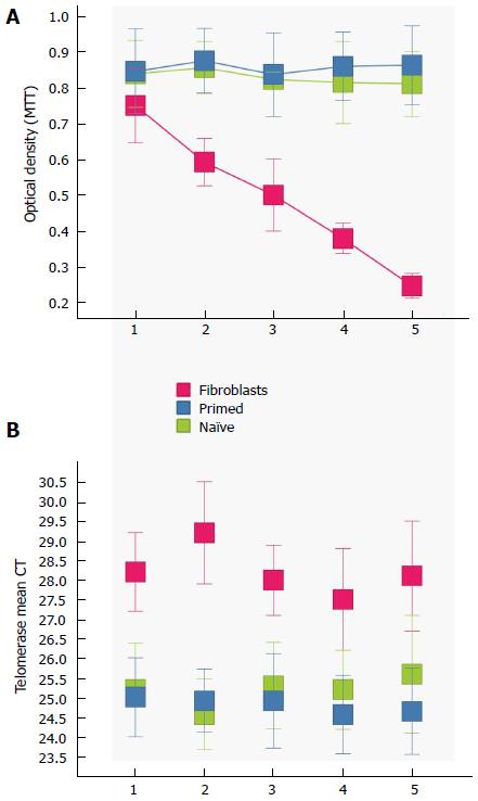
Figure 3 Proliferation and telomerase.
Time course of self-renewal and proliferation of stem cells (potential induced pluripotent stem cells-like cells and embryonic stem cells) relative to control fibroblast (red) as measured by the MTT [3-(4,5-Dimethylthiazol-2-yl)-2,5-diphenilytetrazolium bromide] assay (read at 570 nm). mESCs (blue) and primed mESCs (green) exhibit similar patterns of proliferation, while fibroblast proliferation diminishes as time passes. Telomerase activity was greatly increased (lower mean CT) in both mESCs and primed mESCs over control fibroblast cells. Error bars, SEM (n = 5 independent replicates for both MTT and telomerase data). mESCs: Mouse embryonic stem cells; CT: Cycle threshold.
- Citation: Rossello RA, Pfenning A, Howard JT, Hochgeschwender U. Characterization and genetic manipulation of primed stem cells into a functional naïve state with ESRRB. World J Stem Cells 2016; 8(10): 355-366
- URL: https://www.wjgnet.com/1948-0210/full/v8/i10/355.htm
- DOI: https://dx.doi.org/10.4252/wjsc.v8.i10.355


