Copyright
©The Author(s) 2016.
World J Stem Cells. Oct 26, 2016; 8(10): 342-354
Published online Oct 26, 2016. doi: 10.4252/wjsc.v8.i10.342
Published online Oct 26, 2016. doi: 10.4252/wjsc.v8.i10.342
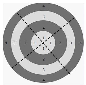
Figure 2 The scheme for photographic fields of the bottom of the culture dish.
The numbers mark the concentric regions: The central region (1); the area near the central region (2); the area near the fringe region (3); the fringe region (4). The dotted lines divide the bottom into four sectors. Each sector comprises four photographic fields; thus, overall, there are 16 photographed fields for each culture dish.
- Citation: Emelyanov AN, Borisova MV, Kiryanova VV. Model acupuncture point: Bone marrow-derived stromal stem cells are moved by a weak electromagnetic field. World J Stem Cells 2016; 8(10): 342-354
- URL: https://www.wjgnet.com/1948-0210/full/v8/i10/342.htm
- DOI: https://dx.doi.org/10.4252/wjsc.v8.i10.342


