Copyright
©The Author(s) 2015.
World J Stem Cells. Sep 26, 2015; 7(8): 1127-1136
Published online Sep 26, 2015. doi: 10.4252/wjsc.v7.i8.1127
Published online Sep 26, 2015. doi: 10.4252/wjsc.v7.i8.1127
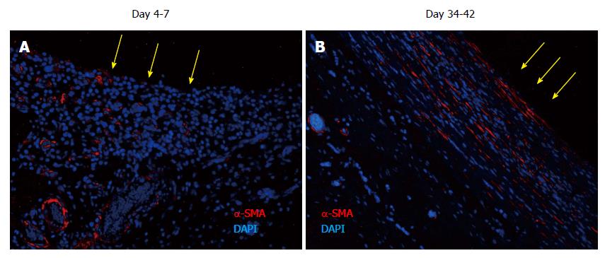
Figure 7 Tissue patch at various times immune-stained for ↑-smooth muscle antigen to determine the organization of the extracellular matrix in the patch.
At day 4-7, α-SMA was found to be associated with blood vessels as expected, but little extracellular α-SMA was observed at these times. At later time points there was more and more of α-SMA observed in the extracellular areas (not shown), which by day 35-42 had stratified and compacted itself in parallel to the length of the implant, consistent with the tissue being tougher and fibrous to touch. Sections were counterstained with DAPI to counterstain nuclei blue. Side in contact with the inert body is shown by yellow arrows. α-SMA: α-smooth muscle antigen; DAPI: 4',6-diamidino-2-phenylindole.
- Citation: Garcia-Gomez I, Gudehithlu KP, Arruda JAL, Singh AK. Autologous tissue patch rich in stem cells created in the subcutaneous tissue. World J Stem Cells 2015; 7(8): 1127-1136
- URL: https://www.wjgnet.com/1948-0210/full/v7/i8/1127.htm
- DOI: https://dx.doi.org/10.4252/wjsc.v7.i8.1127


