Copyright
©The Author(s) 2015.
World J Stem Cells. Aug 26, 2015; 7(7): 1064-1077
Published online Aug 26, 2015. doi: 10.4252/wjsc.v7.i7.1064
Published online Aug 26, 2015. doi: 10.4252/wjsc.v7.i7.1064
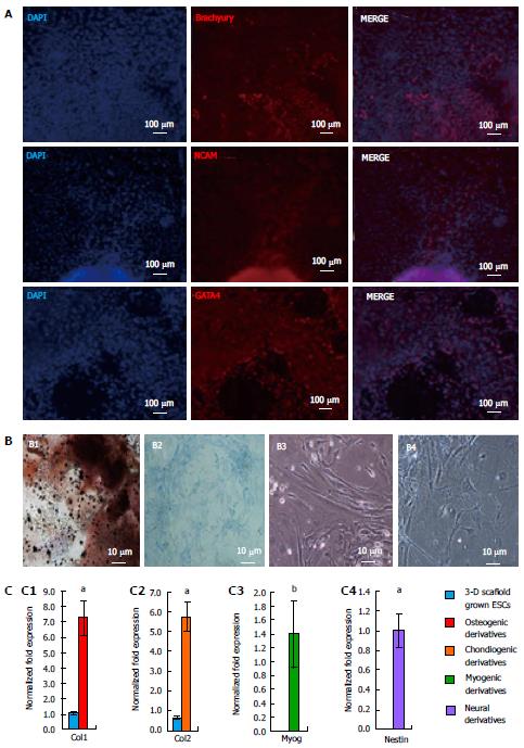
Figure 7 Differentiation potential of three-dimensional grown embryonic stem cells.
A: Embryoid bodies derived from ESCs grown in the 3-D scaffolds were allowed to spontaneously differentiate and analyzed by immunofluorescence staining with antibodies against all three germ layers. Confocal images depicted the presence of Brachyury, NCAM, and GATA4 protein expression, showing the potential of ESCs to differentiate into mesoderm, ectoderm, and endoderm, respectively; B: 3-D scaffold grown ESCs were also directed to differentiate via EB formation and selective induction media. Shown are light micrographs depicting cell morphology of ESC derivatives, osteogenic, chondrogenic, myogenic, and neural (B1, B2, B3, and B4 respectively). Osteogenic and chondrogenic derivatives were cultured for 4 wk and analyzed by von Kossa, and alcian blue staining, respectively. Osteogenic cell-specific ECM stained dark brown for calcium deposition (B1). Proteoglycan produced by chondrogenic derivatives stained blue; C: Expression of cell-specific markers in differentiated ESC derivatives expressed Col1 (C1), Col2 (C2), Myog (C3), and Nestin (C4) markers for osteogenic, chondrogenic, myogenic and neural cells, respectively. Results represent the fold expression ± SE (n = 3) normalized to reference genes Gapdh and β-Actin where a significant (aP < 0.05 and bP < 0.01) increase of marker expression was compared to initial 2-D grown ESCs (day 0), using one-way ANOVA with the non-equal variance hypothesis. ESCs: Embryonic stem cells; ANOVA: Analysis of variance..
- Citation: McKee C, Perez-Cruet M, Chavez F, Chaudhry GR. Simplified three-dimensional culture system for long-term expansion of embryonic stem cells. World J Stem Cells 2015; 7(7): 1064-1077
- URL: https://www.wjgnet.com/1948-0210/full/v7/i7/1064.htm
- DOI: https://dx.doi.org/10.4252/wjsc.v7.i7.1064


