Copyright
©The Author(s) 2015.
World J Stem Cells. Aug 26, 2015; 7(7): 1064-1077
Published online Aug 26, 2015. doi: 10.4252/wjsc.v7.i7.1064
Published online Aug 26, 2015. doi: 10.4252/wjsc.v7.i7.1064
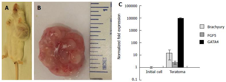
Figure 6 Three-dimensional grown embryonic stem cells formed teratomas in severe combined immunodeficient-beige mice.
Explanted teratoma tissues were analyzed for the expression of three germ layer markers. A: Gross images of tumor growth resulting from injection of 3-D grown ESCs. Tumor growth was observed in all mice injected (n = 3); B: Explanted tumor at 4 wk showed encapsulated, lobular and well-circumscribed gross morphology consistent with teratoma growth; C: Expression of germ layer markers, Brachyury, FGF5, and GATA4 representing mesoderm, ectoderm, and endoderm was analyzed by qRT-PCR. Results of tumor explants were expressed as fold expression ± SE (n = 3) normalized to reference genes Gapdh and β-Actin, and compared to initial cells injected in vivo. ESCs: Embryonic stem cells; qRT-PCR: Quantitative real time polymerase chain reaction..
- Citation: McKee C, Perez-Cruet M, Chavez F, Chaudhry GR. Simplified three-dimensional culture system for long-term expansion of embryonic stem cells. World J Stem Cells 2015; 7(7): 1064-1077
- URL: https://www.wjgnet.com/1948-0210/full/v7/i7/1064.htm
- DOI: https://dx.doi.org/10.4252/wjsc.v7.i7.1064


