Copyright
©The Author(s) 2015.
World J Stem Cells. Jul 26, 2015; 7(6): 956-975
Published online Jul 26, 2015. doi: 10.4252/wjsc.v7.i6.956
Published online Jul 26, 2015. doi: 10.4252/wjsc.v7.i6.956
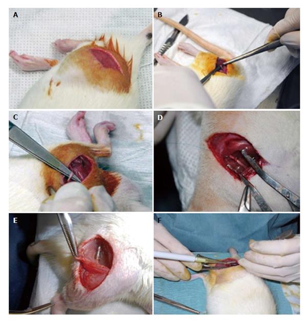
Figure 3 Surgery of the rat sciatic nerve neurotmesis injury model.
Under deep anesthesia the right sciatic nerve was exposed unilaterally through a skin incision extending from the greater trochanter to the mid-half distally followed by a muscle splitting incision (A). After nerve mobilization (B), a transection injury was performed (neurotmesis) using a straight microsurgical scissors (C). In group 5 (End-to-End) after neurotmesis, immediate cooptation with 7/0 monofilament polypropylene suture was performed (D). Implantation of the tube-guide in the 10 mm gap (E). Local application of the mesenchymal stem cells (MSCs), the MSCs suspension filled the polyvinyl alcohol (PVA)-carbon nanotubes (CNTs) tube-guide lumen (group 6: PVA-CNTs-MSCs) (F).
- Citation: Ribeiro J, Pereira T, Caseiro AR, Armada-da-Silva P, Pires I, Prada J, Amorim I, Amado S, França M, Gonçalves C, Lopes MA, Santos JD, Silva DM, Geuna S, Luís AL, Maurício AC. Evaluation of biodegradable electric conductive tube-guides and mesenchymal stem cells. World J Stem Cells 2015; 7(6): 956-975
- URL: https://www.wjgnet.com/1948-0210/full/v7/i6/956.htm
- DOI: https://dx.doi.org/10.4252/wjsc.v7.i6.956


