Copyright
©The Author(s) 2015.
World J Stem Cells. Jul 26, 2015; 7(6): 922-944
Published online Jul 26, 2015. doi: 10.4252/wjsc.v7.i6.922
Published online Jul 26, 2015. doi: 10.4252/wjsc.v7.i6.922
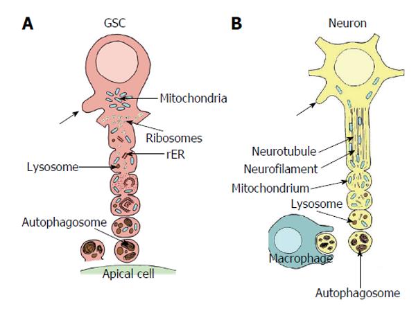
Figure 7 Comparison of autotomizing germline stem cell projection with autodestruction of an injured axon.
A: Sequence of vesicle formation of a GSC projection in Oncopeltus fasciatus. The area of projection formation is characterized by an accumulation of mitochondria (blue). At the base of the projection ribosomes form small clusters (yellow), mitochondria are infrequent. Rough endoplasmic reticulum (rER, orange) and lysosomes (brown) are scarce. With progressing vesicle segregation mitochondria become more frequent and swollen, ribosomes form no longer clusters, rER becomes more prominent and lysosomal bodies increase. Segregated vesicles show many autophagosomes and myelin bodies. They accumulate at the surface of the apical cells (green) and disintegrate. Arrows point to newly sprouting projections (adapted from Dorn et al[60]); B: Sequence of progressive axonal fragmentation after injury. First neurotubules and neurofilaments break down. Then mitochondria accumulate and lysosomes (brown) become more abundant. Finally vesicles which mainly includ autophagosomes and myelin bodies are segregated and taken up by macrophages. Arrows points to newly sprouting axon (adapted from Lingor et al[155]; Beirowski et al[156]; Kerschensteiner et al[157]). GSC: Germline stem cell.
- Citation: Dorn DC, Dorn A. Stem cell autotomy and niche interaction in different systems. World J Stem Cells 2015; 7(6): 922-944
- URL: https://www.wjgnet.com/1948-0210/full/v7/i6/922.htm
- DOI: https://dx.doi.org/10.4252/wjsc.v7.i6.922


