Copyright
©The Author(s) 2015.
World J Stem Cells. Jul 26, 2015; 7(6): 922-944
Published online Jul 26, 2015. doi: 10.4252/wjsc.v7.i6.922
Published online Jul 26, 2015. doi: 10.4252/wjsc.v7.i6.922
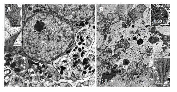
Figure 6 Structural relationships between apical cells and projections and autotomized vesicles of germline stem cells in Oncopeltus fasciatus and Lymantria dispar.
A: Electron micrographs. Oncopeltus fasciatus. Vesicles that are segregated from GSC projections accumulate at the surface of ACs. The vesicles show signs of degeneration. Mitochondria are abundant and appear electron dense. (From Schmidt et al[65]). Upper inset: Intracellular granules in autotomizing GSC projections. Lower inset: Extracellular granules (arrow) between autotomizing GSC projections; B: Electron micrograph. Lymantria dispar. Numerous autotomized GSC projections protrude deeply into the AC (long arrows). Some of the segregated vesicles were apparently taken up by the AC and are being digested (arrow heads). Double arrow points to extracellular granules between GSC vesicles and the AC. Upper inset: Extracellular granules at higher magnification. Lower inset: Tubular indentations into the AC include electron dense material. (From Klein[66]). GSCs: Germline stem cells; AC: Apical cell.
- Citation: Dorn DC, Dorn A. Stem cell autotomy and niche interaction in different systems. World J Stem Cells 2015; 7(6): 922-944
- URL: https://www.wjgnet.com/1948-0210/full/v7/i6/922.htm
- DOI: https://dx.doi.org/10.4252/wjsc.v7.i6.922


