Copyright
©The Author(s) 2015.
World J Stem Cells. Jun 26, 2015; 7(5): 883-893
Published online Jun 26, 2015. doi: 10.4252/wjsc.v7.i5.883
Published online Jun 26, 2015. doi: 10.4252/wjsc.v7.i5.883
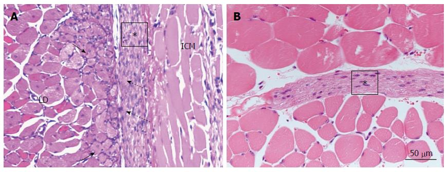
Figure 7 Histology of transplanted domains at 1 wk after transplantation.
A: In G1 NaOH(+), HJV-E(+), Cardiomyocytes(+), myoblasts (dotted long arrows) are marked in the latissimus dorsi muscle (LD). The cardiomyocyte sheet (asterisk) adheres to the myoblast layer at various points (short arrows); B: There are no myoblasts, and no adhesion of the cardiomyocyte sheet (asterisk) to the skeletal muscles in G3 NaOH(-), HJV-E(+), Cardiomyocytes(+). The interspace between the cardiomyocyte sheet and the skeletal muscle fibers is wider. ICM: Intercostal muscle.
- Citation: Takahashi Y, Tomotsune D, Takizawa S, Yue F, Nagai M, Yokoyama T, Hirashima K, Sasaki K. New model for cardiomyocyte sheet transplantation using a virus-cell fusion technique. World J Stem Cells 2015; 7(5): 883-893
- URL: https://www.wjgnet.com/1948-0210/full/v7/i5/883.htm
- DOI: https://dx.doi.org/10.4252/wjsc.v7.i5.883


