Copyright
©The Author(s) 2015.
World J Stem Cells. Jun 26, 2015; 7(5): 883-893
Published online Jun 26, 2015. doi: 10.4252/wjsc.v7.i5.883
Published online Jun 26, 2015. doi: 10.4252/wjsc.v7.i5.883
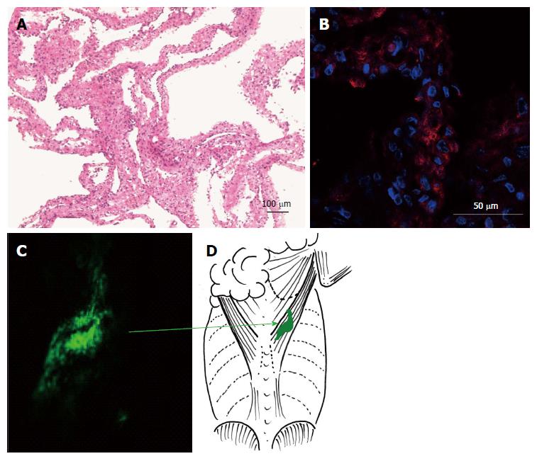
Figure 3 Representative cardiomyocyte sheets.
A: HE staining of a cardiomyocyte sheet separated from the dish; B: The sheets contain cardiac troponin-positive cells (red); C: At 2 wk after transplantation, the DiO-stained sheet (green) occupies the same position as the primary transplanted site (green arrow); D: Schematic diagram showing the transplanted area under the latissimus dorsi muscle.
- Citation: Takahashi Y, Tomotsune D, Takizawa S, Yue F, Nagai M, Yokoyama T, Hirashima K, Sasaki K. New model for cardiomyocyte sheet transplantation using a virus-cell fusion technique. World J Stem Cells 2015; 7(5): 883-893
- URL: https://www.wjgnet.com/1948-0210/full/v7/i5/883.htm
- DOI: https://dx.doi.org/10.4252/wjsc.v7.i5.883


