Copyright
©The Author(s) 2015.
World J Stem Cells. Jun 26, 2015; 7(5): 873-882
Published online Jun 26, 2015. doi: 10.4252/wjsc.v7.i5.873
Published online Jun 26, 2015. doi: 10.4252/wjsc.v7.i5.873
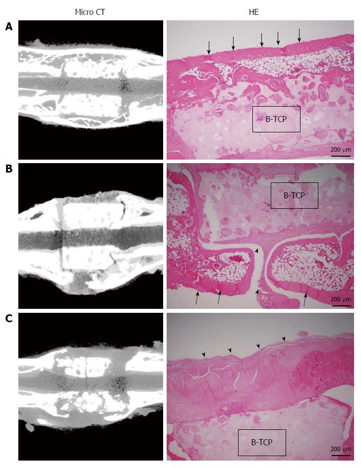
Figure 4 Micro-computed tomography images and hematoxylin and eosin-stained sections at 8 wk post-implantation.
A: BMSC/TCP/Sheet constructs with continuous bone formation to host bone. Both micro-computed tomography (CT) and histology showed bridging bone formation over the TCP to host bone (arrows); B: BMSC/TCP/Sheet constructs with segmental bone formation over the TCP. Soft tissue interposition was observed between the TCP and the callus (arrowheads); C: BMSC/TCP constructs implanted into femurs. The TCP cylinders of the BMSC/TCP constructs were broken. Arrowheads indicate soft tissue interposition between the TCP and the autologous femur. BMSCs: Bone marrow stromal cells; TCP: Tricalcium phosphate.
- Citation: Ueha T, Akahane M, Shimizu T, Uchihara Y, Morita Y, Nitta N, Kido A, Inagaki Y, Kawate K, Tanaka Y. Utility of tricalcium phosphate and osteogenic matrix cell sheet constructs for bone defect reconstruction. World J Stem Cells 2015; 7(5): 873-882
- URL: https://www.wjgnet.com/1948-0210/full/v7/i5/873.htm
- DOI: https://dx.doi.org/10.4252/wjsc.v7.i5.873


