Copyright
©The Author(s) 2015.
World J Stem Cells. May 26, 2015; 7(4): 776-788
Published online May 26, 2015. doi: 10.4252/wjsc.v7.i4.776
Published online May 26, 2015. doi: 10.4252/wjsc.v7.i4.776
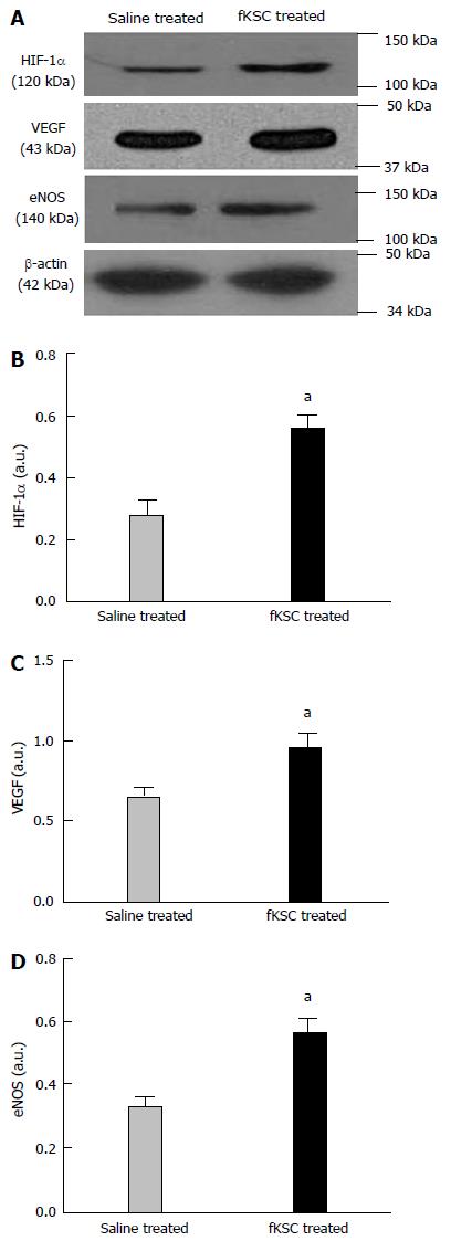
Figure 8 Early angiogenic effect of administered fetal kidney stem cells in cisplatin injured kidney.
Representative immunoblots showing the expression of hypoxia-inducible factor (HIF)-1α, vascular endothelial growth factor (VEGF) and endothelial nitric oxide synthase (eNOS) in saline and fetal kidney stem cells (fKSC) treated groups on day 3 after fKSC therapy (A-D). Bar diagrams showing densitometric quantification of the expression of HIF-1α (B), VEGF (C) and eNOS (D). Comparative gene expression ratio was calculated by referring each gene to β-actin as an internal control. Densitometric analysis applied for comparison of relative protein expression and represented in densitometric arbitrary units (a. u.). Values expressed mean ± SE. aP < 0.05 for fKSC vs saline treated group.
- Citation: Gupta AK, Jadhav SH, Tripathy NK, Nityanand S. Fetal kidney stem cells ameliorate cisplatin induced acute renal failure and promote renal angiogenesis. World J Stem Cells 2015; 7(4): 776-788
- URL: https://www.wjgnet.com/1948-0210/full/v7/i4/776.htm
- DOI: https://dx.doi.org/10.4252/wjsc.v7.i4.776


