Copyright
©The Author(s) 2015.
World J Stem Cells. May 26, 2015; 7(4): 700-710
Published online May 26, 2015. doi: 10.4252/wjsc.v7.i4.700
Published online May 26, 2015. doi: 10.4252/wjsc.v7.i4.700
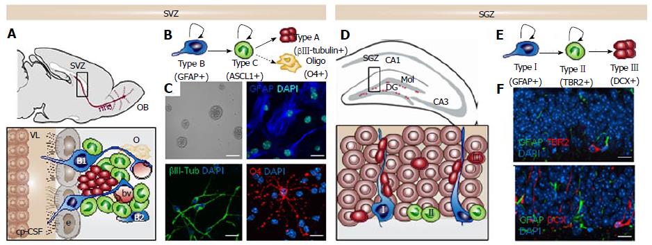
Figure 1 The neurogenic niches in the adult murine mammalian brain.
A: Sagittal view showing the adult mouse subventricular zone (SVZ) and the migrating neuroblasts (red) reaching the olfactory bulb (OB) through the rostral migratory stream (rms). Enlarged view of SVZ: type B1 stem cells (blue) express the astrocyte marker glial fibrillary acidic protein (GFAP) and contact the ventricle with a thin process extended between the ependymal cells (e; gray); type B2 stem cells (blue) contacting the brain parenchyma; transit amplyfing progenitors (TAP) or type C cells (green) express the achaete-scute homolog 1 (ASCL1) transcription factor and give rise to type A cells (red) that migrate through the rostral migratory stream (rms). Dividing stem cells and their TAP progeny are tightly opposed to blood vessels (bv); B: Schematic drawing showing the lineage progression in the SVZ; C: SVZ neural stem cell (NSC) cultures in self-renewal (neurosphere formation) and differentiation. The astrocyte marker GFAP in blue, the neuronal marker βIII-tubulin in green and the oligodendrocyte marker O4 in red; The Choroid plexus-cerebrospinal fluid system (cp-CSF) is shown. D: Coronal view showing the adult mouse subgranular zone (SGZ) and the newborn neurons (red) being integrated in the granular cell layer (gr). Enlarged view of the dentate gyrus (DG): Type I stem cells (blue) are GFAP+ and show a radial single prolongation through the granular layer; Type II precursors give rise to neuronal lineage-restricted progenitors type III cells (red) that differentiate into neurons in the granular layer; E: Schematic drawing showing the lineage progression in the SGZ; F: Confocal images showing immunostaining in the DG for the astrocyte marker GFAP in green, for the progenitor precursor marker T-box brain protein 2 (TBR2) in red and for the neuronal precursor marker Doublecortin (DCX) in red. DAPI is used to stain DNA. Scale bar in C: Top left panel 100 μm, rest 10 μm; In f: 10 μm.
- Citation: Montalbán-Loro R, Domingo-Muelas A, Bizy A, Ferrón SR. Epigenetic regulation of stemness maintenance in the neurogenic niches. World J Stem Cells 2015; 7(4): 700-710
- URL: https://www.wjgnet.com/1948-0210/full/v7/i4/700.htm
- DOI: https://dx.doi.org/10.4252/wjsc.v7.i4.700


