Copyright
©The Author(s) 2015.
World J Stem Cells. Nov 26, 2015; 7(10): 1215-1221
Published online Nov 26, 2015. doi: 10.4252/wjsc.v7.i10.1215
Published online Nov 26, 2015. doi: 10.4252/wjsc.v7.i10.1215
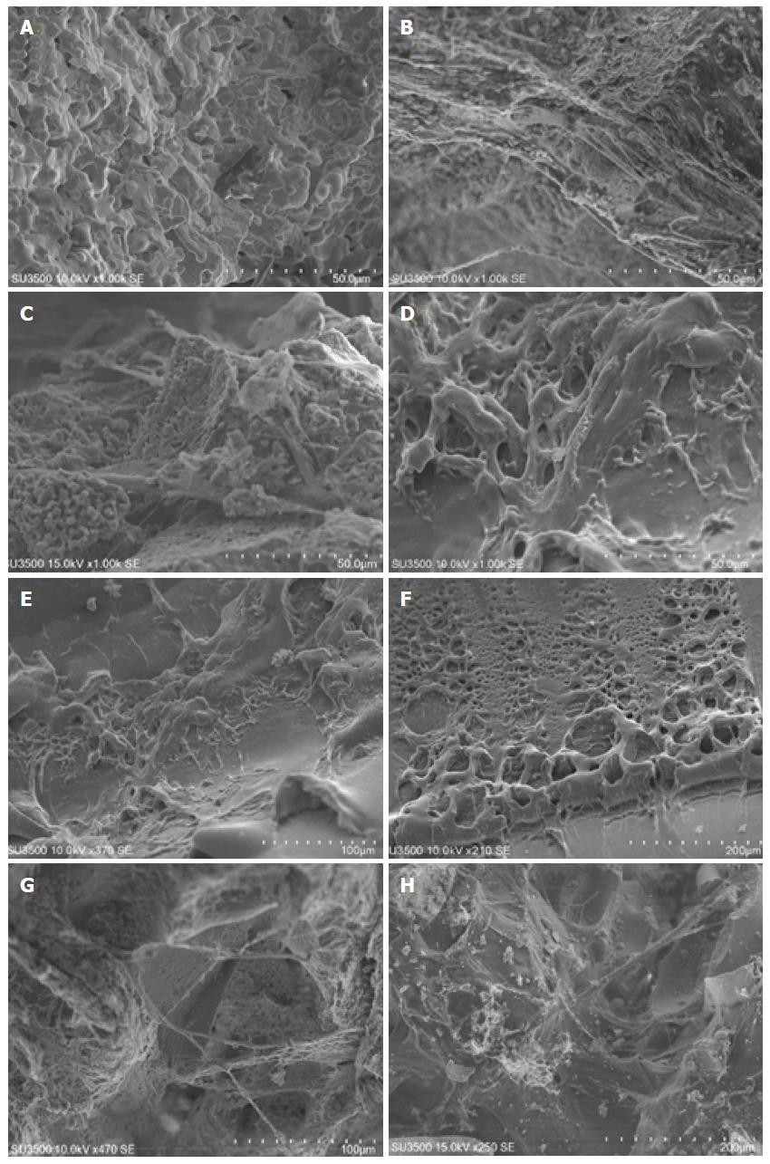
Figure 1 Scanning electron microscopy microphotographs of human dental pulp stem cells attachment and morphology on four different scaffolds.
A: SureOss (Allograft); B: Cerabone (Xenograft); C: OSTEON II Collagen (Composite); D: PLLA (Synthetic); E and F: PLLA at lower magnification; G and H: Cerabone at lower magnification. Note that cells were covered almost the entire scaffold surface of PLLA (D-F). Also note that attached cells on PLLA surface showed fibroblastic morphology (D-F), whereas cells on Cerabone and OSTEON II Collagen demonstrated osteoblasic phenotype (B, C, G, H).
-
Citation: Khojasteh A, Motamedian SR, Rad MR, Shahriari MH, Nadjmi N. Polymeric
vs hydroxyapatite-based scaffolds on dental pulp stem cell proliferation and differentiation. World J Stem Cells 2015; 7(10): 1215-1221 - URL: https://www.wjgnet.com/1948-0210/full/v7/i10/1215.htm
- DOI: https://dx.doi.org/10.4252/wjsc.v7.i10.1215


