Copyright
©2014 Baishideng Publishing Group Inc.
World J Stem Cells. Nov 26, 2014; 6(5): 540-551
Published online Nov 26, 2014. doi: 10.4252/wjsc.v6.i5.540
Published online Nov 26, 2014. doi: 10.4252/wjsc.v6.i5.540
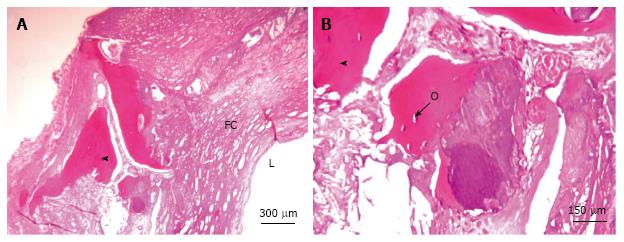
Figure 1 Carotid artery calcifications, hematoxylin and eosin staining.
A: Sheet-like calcifications; B: Osteocytes are visible within the bone lacunae-like mature structure in development with lamellar bone. L: Lumen; FC: Fibrous cap; Arrowhead: Ossification; O: Osteocytes.
- Citation: Vasuri F, Fittipaldi S, Pasquinelli G. Arterial calcification: Finger-pointing at resident and circulating stem cells. World J Stem Cells 2014; 6(5): 540-551
- URL: https://www.wjgnet.com/1948-0210/full/v6/i5/540.htm
- DOI: https://dx.doi.org/10.4252/wjsc.v6.i5.540


