Copyright
©2014 Baishideng Publishing Group Inc.
World J Stem Cells. Nov 26, 2014; 6(5): 511-525
Published online Nov 26, 2014. doi: 10.4252/wjsc.v6.i5.511
Published online Nov 26, 2014. doi: 10.4252/wjsc.v6.i5.511
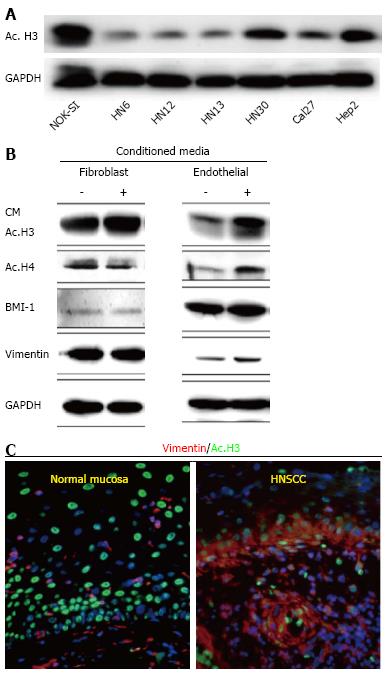
Figure 1 Data represents acetylation status of histone 3 in Head and Neck Squamous Cell Carcinoma by Giudice et al[151].
A: Tumor cells present hypoacetylation of histone 3 (ac.H3) in a panel of Head and Neck Squamous Cell Carcinoma (HNSCC) compared to control cells (NOK-SI); B: Endothelial cell-secreted factors are capable of inducing ac.H3 while fibroblast cell-secreted factors cannot. Also, endothelial cell-secreted factors induce increased expression of BMI-1 and vimentin compared to the fibroblast counterpart; C: Representative examples of human samples of normal oral mucosa and HNSCC. Note acetylated tumor cells (Ac. H3-FITC) with high levels of the epithelial-mesenchymal transition marker vimentin (TRICT) are localized at the invasion front of HNSCC (arrow). Normal mucosa display acetylated cells distributed throughout the epidermis but do not express vimentin. ac. H3: Acetyl histone 3; GAPDH: Glyceraldehyde-3-phosphate dehydrogenase; CM: Conditioned Media; BMI-1: B lymphoma Mo-MLV insertion region 1 homolog; NOK-SI: Normal oral epithelial keratinocytes.
- Citation: Le JM, Squarize CH, Castilho RM. Histone modifications: Targeting head and neck cancer stem cells. World J Stem Cells 2014; 6(5): 511-525
- URL: https://www.wjgnet.com/1948-0210/full/v6/i5/511.htm
- DOI: https://dx.doi.org/10.4252/wjsc.v6.i5.511


