Copyright
©2014 Baishideng Publishing Group Co.
World J Stem Cells. Apr 26, 2014; 6(2): 120-133
Published online Apr 26, 2014. doi: 10.4252/wjsc.v6.i2.120
Published online Apr 26, 2014. doi: 10.4252/wjsc.v6.i2.120
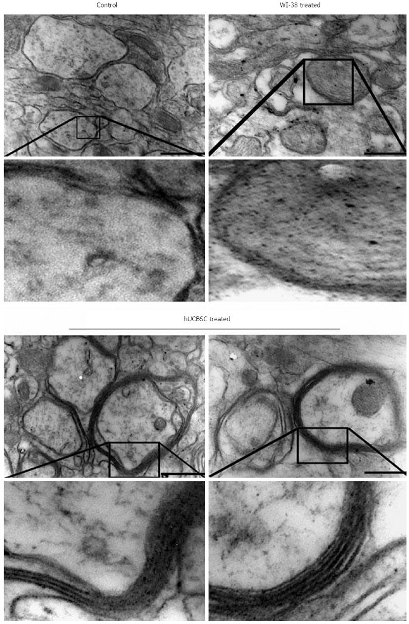
Figure 1 Transmission electron micrographs of shiverer mice brain showing thin and fragmented myelin around the axons in control and WI-38- implanted mice.
In contrast, human umbilical cord blood-derived mesenchymal stem cells-treated shiverer brains showing myelin with several layers. Images are representatives of the several sections obtained from 3 different animals (n = 3). Scale bar = 33000. Stem Cells Dev 2011; 20: 881-891.
- Citation: Dasari VR, Veeravalli KK, Dinh DH. Mesenchymal stem cells in the treatment of spinal cord injuries: A review. World J Stem Cells 2014; 6(2): 120-133
- URL: https://www.wjgnet.com/1948-0210/full/v6/i2/120.htm
- DOI: https://dx.doi.org/10.4252/wjsc.v6.i2.120


