Copyright
©2014 Baishideng Publishing Group Co.
World J Stem Cells. Jan 26, 2014; 6(1): 33-42
Published online Jan 26, 2014. doi: 10.4252/wjsc.v6.i1.33
Published online Jan 26, 2014. doi: 10.4252/wjsc.v6.i1.33
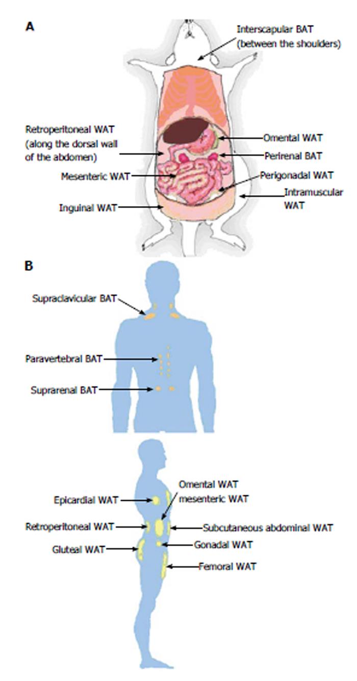
Figure 1 Locations of adipose tissue depots in a mouse (A) and an adult human (B).
A: Subcutaneous (inguinal and intramuscular), visceral (mesenteric, omental, perigonadal and retroperitoneal) and brown (interscapular and perirenal) adipose tissue depots are shown in a mouse model; B: Subcutaneous (abdominal, femoral and gluteal), visceral (epicardial, gonadal, mesenteric, omental and retroperitoneal) and brown (paravertebral, supraclavicular and suprarenal) adipose tissue depots are shown in a human model. WAT: White adipose tissue; BAT: Brown adipose tissue.
- Citation: Park A, Kim WK, Bae KH. Distinction of white, beige and brown adipocytes derived from mesenchymal stem cells. World J Stem Cells 2014; 6(1): 33-42
- URL: https://www.wjgnet.com/1948-0210/full/v6/i1/33.htm
- DOI: https://dx.doi.org/10.4252/wjsc.v6.i1.33


