Copyright
©2013 Baishideng Publishing Group Co.
World J Stem Cells. Oct 26, 2013; 5(4): 205-216
Published online Oct 26, 2013. doi: 10.4252/wjsc.v5.i4.205
Published online Oct 26, 2013. doi: 10.4252/wjsc.v5.i4.205
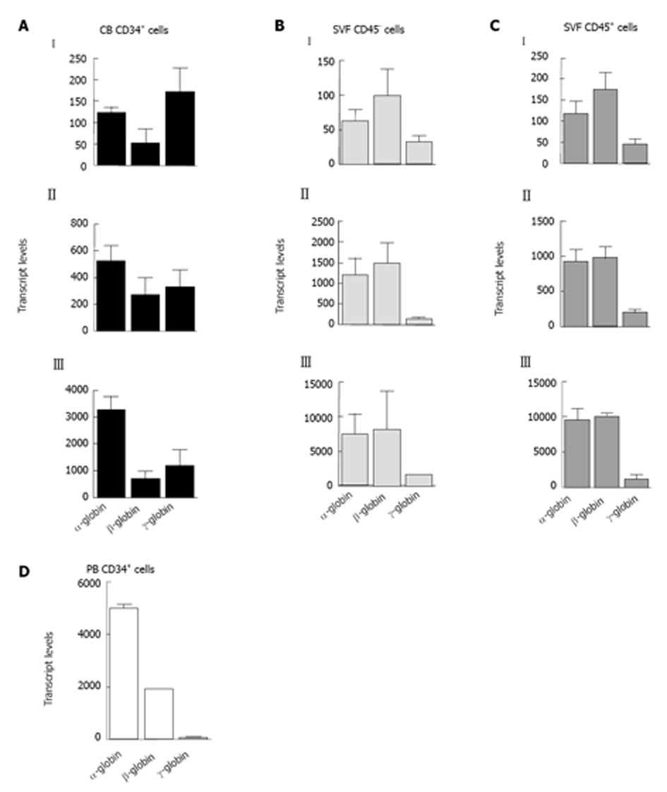
Figure 4 Analysis of globin gene expression in erythroid cells.
CD45+ and CD45- cells isolated from the stromal vascular fraction (SVF) and CD34+ cells from cord blood (CB) or adult peripheral blood (PB) were cultured in a methylcellulose-based medium, and burst-forming units-erythroid -derived erythroid cells were isolated at day 15 of culture to determine globin gene expression by reverse transcription-polymerase chain reaction. The transcripts were normalized to glyceraldehyde-3- phosphate dehydrogenase. Based on the α-globin levels, the values obtained for SVF- and CB-derived cells were placed into three groups (I, II and III). A: CB CD34+ cells, n = 10; B: SVF CD45- cells, n = 17; C: SVF CD45+ cells, n = 17; D: PB CD34+ cells, n = 4. All samples were assayed in duplicate.
- Citation: Navarro A, Carbonell-Uberos F, Marín S, Miñana MD. Human adipose tissue contains erythroid progenitors expressing fetal hemoglobin. World J Stem Cells 2013; 5(4): 205-216
- URL: https://www.wjgnet.com/1948-0210/full/v5/i4/205.htm
- DOI: https://dx.doi.org/10.4252/wjsc.v5.i4.205


