Copyright
©2012 Baishideng.
World J Stem Cells. Aug 26, 2012; 4(8): 80-86
Published online Aug 26, 2012. doi: 10.4252/wjsc.v4.i8.80
Published online Aug 26, 2012. doi: 10.4252/wjsc.v4.i8.80
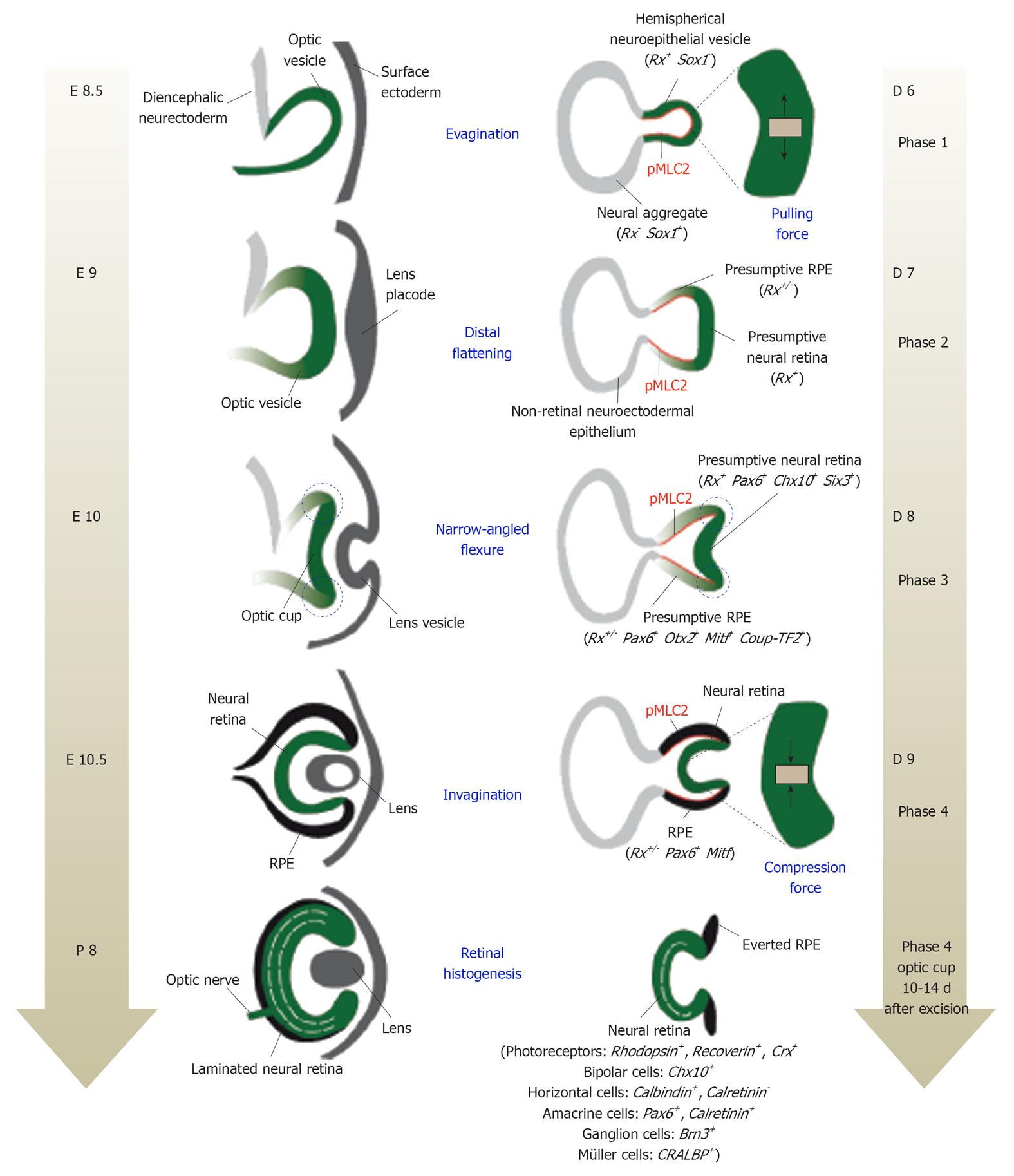
Figure 1 Schematic representation of in vivo (A) and in vitro (B) retinal development.
A: During mouse embryonic development, the Rx-expressing optic vesicle (green) evaginates from the diencephalon at embryonic day 8 (E8). After contacting the surface ectoderm, the outer wall of the optic vesicle invaginates to form the optic cup. Its proximal layer forms the retinal pigmented epithelium (RPE), while the distal layer constitutes the neural retina. Retinal histogenesis and consecutive lamination then occurs. The process is completed postnatally (P0-P11); B: Retinal morphogenesis from mouse embryonic stem cells mimics this whole in vivo process and can be divided into 4 main phases. In phase 1, the Rx-expressing neuroepithelium (green) evaginates from the neural aggregate. In phase 2, its distal portion flattens. During phase 3, the angles at the hinge between the presumptive RPE and the neural retina become acute (dotted blue circles). Finally, a two-walled optic cup is formed during phase 4, following invagination of the retinal neuroepithelium. Pulling and compression forces that contribute to these morphogenetic changes are illustrated with black arrows in the neural retina magnifications. Progressive acquisition of neural retina- and RPE-specific identities is highlighted by the indication of selectively expressed set of genes. From phase 2, both tissues can also be distinguished in terms of mechanical properties, with the RPE (and hinge points) being more rigid than the NR. This correlates with higher levels of Rho-associated protein kinase activity, visualized by the expression of phosphorylated Myosin Light Chain 2 (pMLC2; red outline). Finally, following excision and further culture, phase 4 optic cup recapitulates retinal histogenesis, as observed in vivo. As a minor difference compared to the in vivo situation, synthetic retinas exhibit scattered ganglion cells and fewer cone photoreceptors. Note also that optic cup excision leads to RPE eversion.
- Citation: Colozza G, Locker M, Perron M. Shaping the eye from embryonic stem cells: Biological and medical implications. World J Stem Cells 2012; 4(8): 80-86
- URL: https://www.wjgnet.com/1948-0210/full/v4/i8/80.htm
- DOI: https://dx.doi.org/10.4252/wjsc.v4.i8.80


