Copyright
©2012 Baishideng.
World J Stem Cells. May 26, 2012; 4(5): 35-43
Published online May 26, 2012. doi: 10.4252/wjsc.v4.i5.35
Published online May 26, 2012. doi: 10.4252/wjsc.v4.i5.35
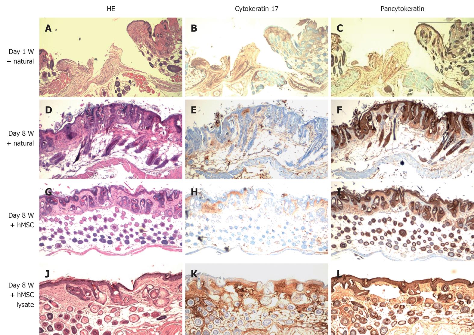
Figure 6 HE (panels A, D, G and J), cytokeratin 17 (panels B, E, H and K) and pancytokeratin staining (panels C, F, I and L) of skin sections.
Figure shows restoration of both dermis and epidermis in skins of mice (nu/nu age: 4-5 wk) treated with human mesenchymal stem cell (hMSC), hMSC lysate and CMC from hMSC; A-C: Wounded skin sections (day 1); D-F: Wounded skin allowed to heal naturally (day 8); G-I: Large numbers of pancytokeratin positive cells were observed in the dermis of hMSC administered wounded skin; J-L: hMSC Lysate injected skin sections. Scale bar: 100 μm.
- Citation: Mishra PJ, Mishra PJ, Banerjee D. Cell-free derivatives from mesenchymal stem cells are effective in wound therapy. World J Stem Cells 2012; 4(5): 35-43
- URL: https://www.wjgnet.com/1948-0210/full/v4/i5/35.htm
- DOI: https://dx.doi.org/10.4252/wjsc.v4.i5.35


